Today’s topic: Marchiafava-Bignami disease.
When the deep neural pathway connecting the left and right sides of the brain begins to break down, the sudden shift in a person’s behavior and motor control can look terrifying.
Doctors call this specific type of neurological crisis Marchiafava-Bignami disease. It is a rare but incredibly severe condition where the central bridge of the brain undergoes rapid degeneration.
Usually triggered by years of severe alcohol misuse coupled with extreme nutritional neglect, it essentially starves brain cells of the basic vitamins they need to function.
Catching this early is everything. Missing the warning signs can lead to permanent cognitive damage within days.
This guide breaks down the latest medical data, clear diagnostic markers, and treatment pathways. It also shows how healthcare teams spot and manage this complex condition to give patients the best chance of recovery.
What Is Marchiafava-Bignami Disease?
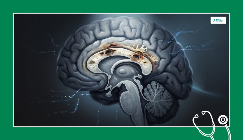
Medical professionals describe the condition as an exceptionally rare neurological disorder defined by the progressive demyelination, necrosis, and atrophy of the corpus callosum.
First identified by Italian pathologists Vittorio Marchiafava and Amico Bignami in 1903, the disease first surfaced historically among individuals consuming specific types of home-reared red wine.
According to modern clinical research, Marchiafava-Bignami disease is not limited by geography or specific beverage types. Instead, profound metabolic and nutritional stress fundamentally drives it.
The primary structural target of this pathology is the central white matter pathway connecting the left and right cerebral hemispheres.
When this bundle of nerve fibers fails, interhemispheric information transfer fails. As a result, a distinct constellation of cognitive, behavioral, and motor deficits emerges.
The Nutritional And Toxic Origins
The exact molecular mechanisms causing this disorder involve a mix of direct alcohol toxicity and severe B-complex vitamin deficiencies. Prolonged alcohol abuse damages the lining of the gastrointestinal tract.
Accordingly, this severely inhibits the body’s ability to absorb essential micronutrients.
As a result, the body lacks adequate thiamine and related B vitamins. Therefore, cellular metabolism inside the brain slows down drastically, leading to high cellular oxidative stress.
This localized metabolic failure specifically targets the oligodendrocytes. These specialized cells maintain the protective myelin sheath.
Vulnerability Of The Corpus Callosum
The corpus callosum has an incredibly high density of myelin and a unique vascular supply. This makes it especially vulnerable to rapid drops in metabolism.
During prolonged malnutrition, the blood-brain barrier in this region becomes highly permeable and unstable.
This localized breakdown allows cytotoxic fluid to accumulate, causing the nerve tissues to swell rapidly. Eventually, they start to suffocate. If you leave this metabolic starvation unchecked, the central layer of the callosum slowly dissolves.
As a result, permanent fluid-filled cystic cavities remain.
Read Also: When The Body Attacks The Senses: How Susac Syndrome Mimics MS And Hijacks Your Senses
Clinical Symptoms And Diagnostic Presentations
The clinical presentation of this illness varies dramatically depending on how quickly the underlying tissue destruction spreads through the central nervous system.
Neurologists generally divide the disorder into clinical types to guide emergency triage and long-term care tracking.
| Clinical Variant | Onset Speed | Dominant Clinical Features | Primary Outcomes |
| Type A (Acute) | Hours to days | Coma, seizures, structural stupor, limb paralysis | High risk of early mortality |
| Type B (Subacute) | Weeks | Variable confusion, behavioral changes, gait ataxia | Potential for partial recovery |
| Type C (Chronic) | Months to years | Progressive dementia, speech deficits, apraxia | Long-term neurological decline |
Cognitive And Behavioral Declines
Patients experiencing the onset of Marchiafava-Bignami disease frequently exhibit sudden and dramatic personality transformations. These mimic acute psychiatric emergencies.
Oftentimes, families report uncharacteristic bouts of aggressive behavior, profound emotional apathy, and severe depression, defying typical behavioral patterns.
As structural damage in the brain spreads, executive functions like working memory, sustained attention, and problem-solving deteriorate completely.
Moreover, clinicians frequently misdiagnose these symptoms as standard alcohol dementia, delaying vital medical interventions.
Neurological And Motor Deficits
Experts say motor deterioration is a major indicator of the condition. Typically, this starts as a wide-based unstable walking pattern known as gait ataxia. As the callosal pathways degrade, patients begin to lose fine motor control.
As a result, they experience severe muscle tremors, localized spasticity, and balance difficulties.
When it comes to advanced cases of Marchiafava-Bignami disease, individuals start to develop split-brain syndrome. This is when the hemispheres function completely out of sync.
How Clinicians Diagnose The Condition
Before modern neuroimaging, doctors accurately diagnosed this illness almost exclusively during post-mortem autopsies.
Today, advanced brain imaging allows medical teams to identify structural changes early enough to design effective treatment plans.
Breakdown Of Primary Diagnostic Tools Used In Modern Hospitals
| Diagnostic Tool | Specific Purpose | Key Clinical Findings |
| MRI (T2 & FLAIR) | Visualizing structural callosal damage | Symmetrical lesions; classic “sandwich sign” |
| Diffusion-Weighted Imaging | Early metabolic tracking | Subtle cellular changes within days of onset |
| Comprehensive Blood Panels | Ruling out metabolic mimics | Liver anomalies, electrolyte imbalances, vitamin drops |
| Spinal Taps & EEGs | Checking alternative causes | Rules out active CNS infections or hidden seizures |
Magnetic Resonance Imaging (MRI) Markers
Modern medical teams use MRI as the definitive tool to identify the classic signatures of Marchiafava-Bignami disease.
T2-weighted and Fluid-Attenuated Inversion Recovery (FLAIR) sequences typically reveal bright, symmetrical lesions stretching across the corpus callosum body.
These images often show a sign known as the “sandwich sign.” Here, the middle layer of the callosum is damaged while the outer edges remain intact.
Diffusion-weighted imaging can detect these subtle cellular changes within just a few days of symptom onset.
Crucial Differential Diagnosis Tests
Because the symptoms overlap heavily with other neurological conditions, doctors must carefully rule out other disorders. This includes diseases such as Wernicke’s encephalopathy and central pontine myelinolysis.
Doctors order comprehensive blood panels immediately to check for liver function anomalies, severe electrolyte imbalances, and systemic vitamin deficiencies.
Doctors also utilize spinal taps and electroencephalograms to rule out active central nervous system infections or hidden seizure patterns. Combining these lab evaluations with precise neuroimaging ensures an accurate diagnosis.
Modern Treatment Protocols And Recovery Expectations
Successful management of Marchiafava-Bignami disease requires an aggressive, multi-layered treatment plan implemented as soon as symptoms appear.
Since tissue death can become irreversible within days, medical teams prioritize rapid metabolic stabilization over lengthy testing windows.
Intravenous Micronutrient Therapy
The absolute foundation of medical management is the immediate delivery of high-dose intravenous thiamine, along with other essential B-complex vitamins. This aggressive replenishment strategy bypasses the damaged digestive system.
Moreover, it supplies critical nutrients directly to struggling brain cells.
When a patient presents with a history of alcoholism and sudden confusion, clinicians routinely start this vitamin regimen before completing MRI scans.
Medical teams also perform spinal taps and electroencephalograms to rule out active central nervous system infections or hidden seizure patterns. As a result, it helps reduce localized tissue swelling and inflammation.
Expectations For Complete Structural Reversibility
A significant question in critical care arises. Can critical care medicine ever completely reverse the callosal brain damage from Marchiafava-Bignami disease?
Full structural recovery is possible only with aggressive treatment.
It should be started during the initial demyelination stage, before tissue death occurs.
Once the disease advances to permanent tissue necrosis or fluid-filled cyst formation, the physical scars in the corpus callosum become permanent features.
Even with permanent scarring, intensive long-term rehabilitation can still help patients recover an impressive amount of daily functionality.
Comprehensive Neuro-Rehabilitation
Once a patient’s condition stabilizes, care shifts to an intensive program of physical, speech, and occupational therapy. These targeted sessions are designed to stimulate neuroplasticity.
This helps the brain to rebuild alternative pathways around the permanent callosal scar tissue.
Strict and lifelong alcohol cessation is absolutely mandatory to protect the fragile central nervous system from further toxic injury.
Regular nutritional counseling and targeted dietary supplements are maintained indefinitely to give the body the tools it needs for long-term nerve repair.
Understanding The Overall Prognosis Of Marchiafava-Bignami Disease
Historically, the long-term outlook for individuals diagnosed with Marchiafava-Bignami disease was incredibly poor.
Additionally, it carried an exceptionally high mortality rate.
However, the rise of accessible MRI scanning and rapid intravenous vitamin therapies has significantly improved patient outcomes.
While some survivors face permanent cognitive deficits, many achieve remarkable levels of physical independence when treated early.
The time gap between symptom onset and the start of vitamin therapy remains the single most critical factor in predicting overall recovery.
| Disclaimer: This article is for informational and educational purposes only and does not substitute for professional medical advice. Reading this content does not create a doctor-patient relationship. Always consult a qualified healthcare provider for medical concerns and never delay seeking professional advice based on what you read here. |
References
- Tian, T. Y., Ruschel, M. A. P., Park, S., & Liang, J. W. (2023, July 24). Marchiafava-Bignami Disease. StatPearls – NCBI Bookshelf.
- Choi, H. J., & Han, S. W. (2025). Marchiafava–Bignami disease. Journal of Neurosonology and Neuroimaging, 17(2), 53–54.