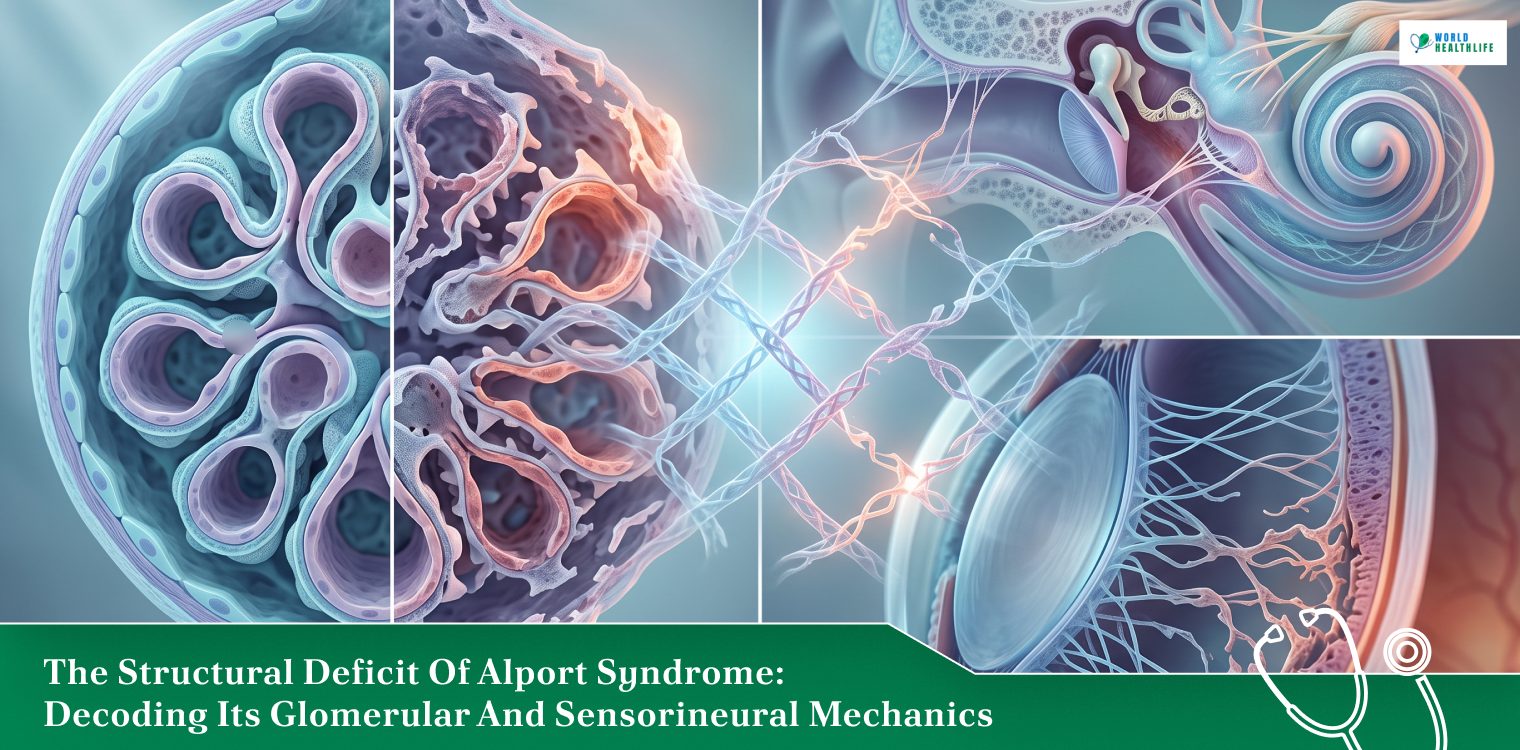
Within the structural framework of the human body, the extracellular matrix acts as the invisible scaffolding that preserves tissue boundaries and cellular integrity.
In specialized organ systems, this structural support relies almost entirely on a highly specific protein network, which is Type IV collagen.
For instance, organ systems such as the filtering units of the kidneys, the delicate chambers of the inner ear, and the refractive layers of the eye have this.
When this molecular mesh network is compromised by genetic variations, multiple organ systems experience a progressive functional breakdown.
When evaluating Alport syndrome, clinicians treat the condition not merely as an isolated kidney disease. Instead, it is a progressive, multi-system basement membrane disorder.
The underlying genetic variations cause a structural failure in the production of collagen alpha chains.
This leads to a steady decline in renal filtration capacity along with sensorineural hearing loss and ocular anomalies.
Managing this complex genetic condition requires looking past basic symptom descriptions.
Navigating the clinical realities of this diagnosis requires an objective look at the precise molecular mechanics of basement membrane degradation.
Thus, we take this into consideration in this guide. Besides, it will also delve deeply into the varying modes of genetic transmission and the modern therapeutic strategies used to delay end-stage renal disease (ESRD).
The Molecular Pathology: The Type IV Collagen Network
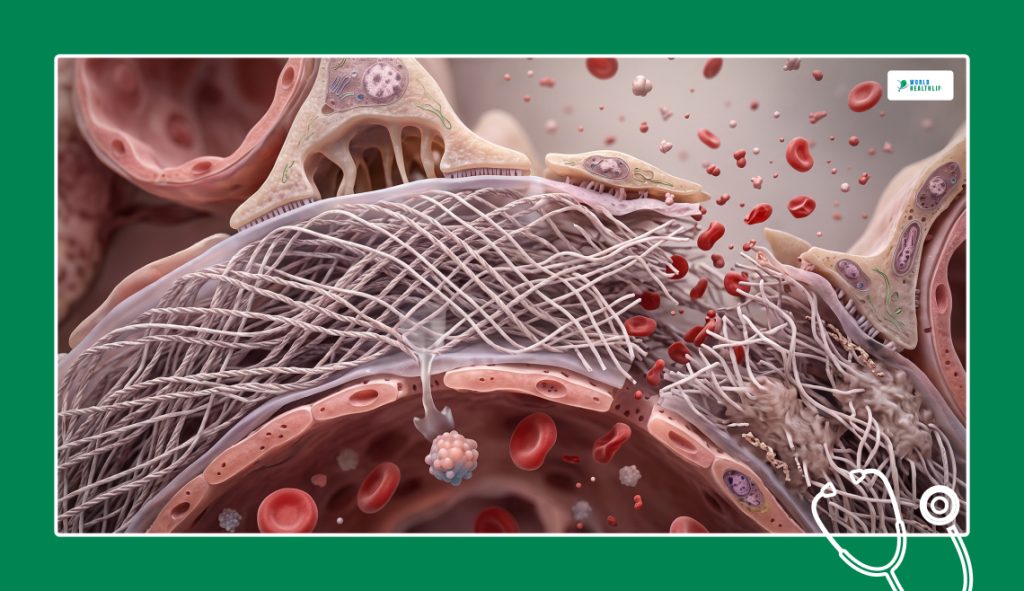
First, before we dive deep into how Alport syndrome unravels tissue function, you have to look directly at the structural layout of the glomerular basement membrane (GBM). This is located inside the kidneys.
In a healthy adult body, the GBM is built from a specialized triple-helix meshwork. This is composed of α3, α4, and α5 Type IV collagen chains.
This dense molecular network forms a tough and selective filtration barrier that allows liquid waste to pass into the urine. On the other hand, it securely keeps vital blood cells and heavy proteins inside the bloodstream.
However, in individuals with this condition, genetic mutations disrupt the synthesis of these advanced alpha chains.
The body cannot produce the mature α3-α4-α5 network. Therefore, it is forced to rely on an embryonic, fragile α1-α1-α2 collagen network instead
This immature replacement is highly vulnerable to ongoing mechanical stress and enzymatic breakdown.
Over time, the continuous high-pressure filtration environment inside the kidney causes the fragile basement membrane to split, thin, and tear.
As the filtration barrier develops dense structural scars, microscopic blood cells and vital proteins begin leaking through the gaps. As a result, it sets off a progressive decline toward total renal failure.
Tracking The Genetic Transmission Pipelines
The clinical severity, onset timeline, and overall progression of the disease are dictated entirely by the specific genetic pathway inherited by the patient.
The condition maps across three primary genetic distribution models.
| Inheritance Mode | Involved Gene Locus | Population Prevalence | Gender-Specific Disease Expression |
| X-Linked Alport Syndrome (XLAS) | COL4A5 gene mutation located on the X chromosome. | Represents roughly 80% to 85% of all diagnosed cases. | Males experience aggressive, rapid progression toward early ESRD; females show variable, often milder symptoms due to random X-inactivation. |
| Autosomal Recessive (ARAS) | Homozygous or compound heterozygous mutations in COL4A3 or COL4A4. | Accounts for approximately 10% to 15% of the clinical pool. | Affects biological males and females with equal severity, frequently causing early-onset renal failure before age thirty. |
| Autosomal Dominant (ADAS) | Heterozygous mutation in either the COL4A3 or COL4A4 genes. | The rarest variant, representing roughly 5% of cases. | Displays a significantly slower, indolent progression profile; kidney failure often delays until late adulthood. |
The Extra-Renal Triad: Hearing And Vision Anomalies
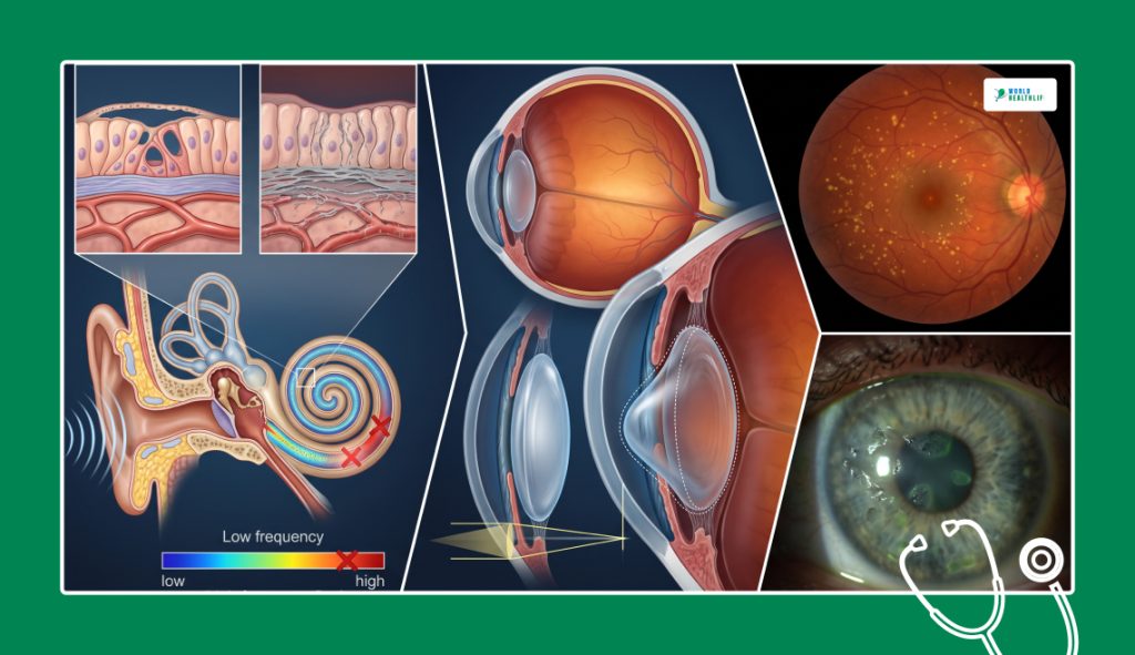
Type IV collagen alpha chains are heavily concentrated inside the delicate sensory mechanisms of the head. Moreover, the structural breakdown extends far past the renal architecture.
The progressive thinning of the basement membrane creates a highly distinct clinical triad of symptoms.
Sensorineural Hearing Loss Mechanics
The inner ear relies on a specialized structural membrane called the stria vascularis. The primary function of this is to maintain the complex fluid balance required to transmit sound waves to the brain.
In patients with X-linked or autosomal recessive forms of the disease, there is structural degradation of the basement membrane inside the cochlea.
Consequently, it causes progressive, bilateral sensorineural hearing loss.
This hearing decline is typically unnoticeable in early childhood. However, it begins manifesting during late childhood or early adolescence, primarily striking high-frequency sound registers first.
Ocular Structural Manifestations
The eye contains multiple clear and high-stress boundaries. These have a heavy reliance on rigid collagen support to maintain optical focus,
The most signature ocular sign of the syndrome is anterior lenticonus. This is a unique condition where the thin capsule wrapping around the eye’s natural lens becomes progressively fragile.
Accordingly, it leads to the lens bulging forward into a conical shape.
This structural shift triggers a progressive, severe myopia (nearsightedness) that requires specialized corrective lenses.
Furthermore, clinicians routinely isolate distinctive dot-and-fleck retinopathy patterns across the retina. That is, along with recurrent corneal erosions that cause temporary eye pain and light sensitivity.
Advanced Endocrinology And Nephrology Diagnostics
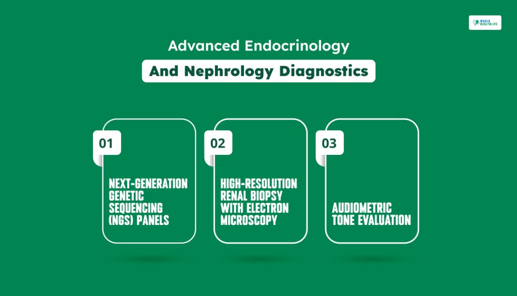
The achievement of a definitive diagnosis for Alport syndrome requires you to move past superficial urine checks.
Moreover, you need to deploy high-precision genetic and structural screening tools.
Next-Generation Genetic Sequencing (NGS) Panels
This tool is considered to be the definitive clinical gold standard.
NGS tracks specific mutations across the COL4A3, COL4A4, and COL4A5 gene chains to confirm the exact inheritance mode. Additionally, the tools help to guide family planning decisions.
High-Resolution Renal Biopsy With Electron Microscopy
If genetic results are unclear, a tissue sample is taken from the kidney.
Under an electron microscope, a classic Alport kidney displays a highly distinct “basket-weave” pattern, characterized by irregular thinning, thickening, and longitudinal splitting of the glomerular basement membrane layers.
Audiometric Tone Evaluation
Regular and high-frequency hearing checks track subtle changes in sensorineural function.
This is before the patient experiences noticeable conversational communication barriers.
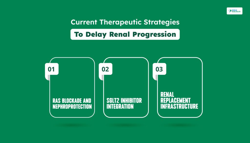
Current Therapeutic Strategies To Delay Renal Progression
There is currently no corrective gene therapy available to rewrite the broken collagen blueprints at the root of the condition.
Conversely, modern nephrology utilizes aggressive pharmacological interventions to prolong kidney life and protect the cardiovascular system.
RAS Blockade And Nephroprotection
The cornerstone of modern disease management is the early deployment of Renin-Angiotensin System (RAS) inhibitors. For instance, Angiotensin-Converting Enzyme (ACE) inhibitors or Angiotensin Receptor Blockers (ARBs).
These medications lower the physical blood pressure inside the filtering units of the kidney. Accordingly, this reduces the mechanical stress placed on the fragile basement membrane.
As per clinical data, starting ACE inhibitor therapy early significantly delays the onset of end-stage renal disease. This is ideally during the microalbuminuria stage before heavy protein loss occurs.
As a result, it adds decades of functional kidney life for many patients.
SGLT2 Inhibitor Integration
Recent clinical protocols have expanded to integrate sodium-glucose cotransporter 2 (SGLT2) inhibitors into the standard nephroprotective toolkit.
Originally designed as diabetes medications, these drugs provide powerful, independent renal protection.
To do so, it alters fluid pressures inside the filtering units and reduce localized tissue inflammation. This offers a secondary line of defense alongside traditional RAS blockades.
Renal Replacement Infrastructure
When a patient eventually transitions into end-stage renal failure, long-term survival requires moving toward automated dialysis options or a kidney transplant.
Fortunately, patients with Alport syndrome generally display excellent long-term post-transplant survival outcomes.
The newly transplanted kidney comes from a donor with healthy and normal collagen genes. Thus, the disease cannot recur inside the new organ.
However, transplant teams must closely monitor the patient’s immune response.
In rare instances, the patient’s immune system may view the normal Type IV collagen in the new kidney as a foreign object. Consequently, it triggers a rare, specialized complication known as anti-GBM nephritis.
Restoring Long-Term Vitality Safely
Deciphering the mechanics of Alport syndrome proves that managing genetic tissue disorders requires a systematic pivot away from reactive symptom care toward proactive, early structural protection.
In addition, the body’s intricate filtration and sensory networks can only maintain their vital rhythms when their foundational basement membranes are actively shielded from high mechanical pressures.
First of all, you need to deploy next-generation genetic sequencing early and utilize targeted RAS blockades. This will help you lower filtration stress.
Then, by coordinating close multidisciplinary care among nephrologists, audiologists, and ophthalmologists, you can manage disease progression.
It takes lifelong clinical consistency and precise, patient-specific tracking. However, it allows individuals to step out of the progressive crossfire.
Therefore, they can maximize their functional kidney longevity, protect their sensory health, and preserve their long-term quality of life.
Disclaimer: The information provided in this article is for general informational purposes only. It does not, and is not intended to, constitute medical or health advice. Please consult a qualified healthcare professional for medical guidance or expert health assistance.