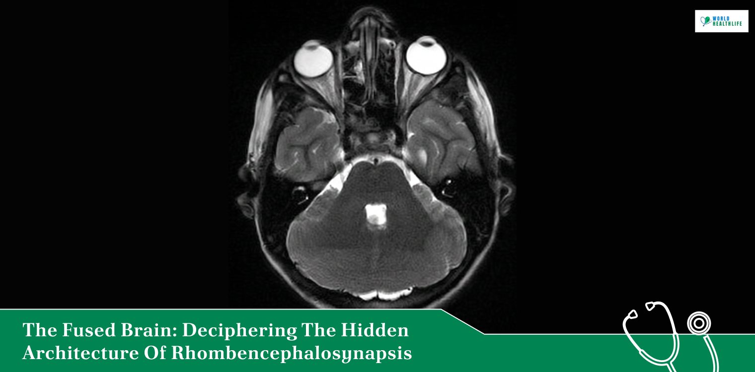
Most people think of the brain as two separate hemispheres that work in harmony. Here, the cerebellum is the control centre which has the responsibility for motor coordination, balance, and fine physical skills.
In the human cerebellum, this split design is completely vital. The left and right cerebellar lobes are supposed to remain distinct, connected only by a central midline bridge known as the vermis.
However, sometimes, a rare developmental error can change the entire blueprint before a child is even born. In the case of Rhombencephalosynapsis, the structural lines separating the hemispheres never form.
Instead, the two halves of the cerebellum fuse together into a single, continuous mass of tissue.
This ultra-rare hindbrain malformation bypasses classic syndromic boundaries. It leaves patients navigating a massive, unpredictable range of physical, visual, and behavioral challenges.
Understanding this condition takes a deep, no-nonsense dive into its core structural defects, its unique radiological signs, and what it actually takes to manage the clinical tracking of symptoms over time.
Defining the Structural Defect: A Failure to Separate
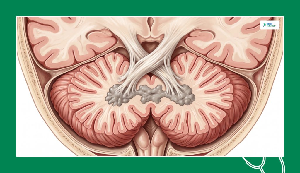
The term Rhombencephalosynapsis describes a highly specific embryological failure that happens very early in gestation. Additionally, it is likely to occur between days 28 and 44 of embryonic development.
The primary pathology centers on the complete or partial agenesis of the cerebellar vermis. Without this central separator, the developing nerve fibers fuse directly across the midline.
Consequently, this structural glitch changes the entire landscape of the posterior fossa.
The condition does not just fuse the outer tissue of the hemispheres. Instead, it forces a deep, structural fusion of the dentate nuclei and the superior cerebellar peduncles.
Something forces these deep white-matter pathways into a single, tangled block. As a result, the brain loses the ability to transmit smooth, coordinated signals down the spinal cord.
Unveiling The Donald Duck Sign On Neuroimaging
Confirming this condition relies heavily on high-resolution magnetic resonance imaging (MRI) scans.
When a neuroradiologist runs a coronal cut or a T2-weighted view on a patient, the structural fusion creates a highly distinct visual pattern. Medical professionals call this the “Donald Duck sign.”
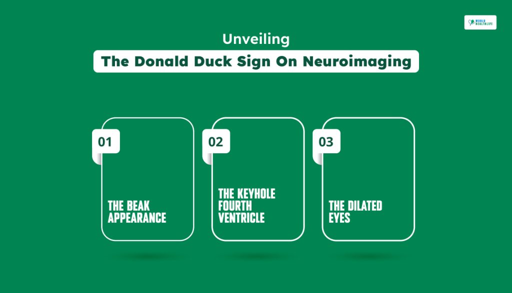
This signature imaging mark is caused by a unique combination of three separate structural shifts occurring side-by-side.
This blockage forces a massive ventriculomegaly, ballooning the lateral and third ventricles into large circular shapes. Furthermore, they look exactly like wide cartoon eyes sitting right above the fused beak on a coronal scan.
Finding this clear visual marker on a brain MRI immediately separates the condition from other common posterior fossa issues.
Issues such as the Dandy-Walker continuum or Joubert syndrome, allowing clinical teams to form a rapid diagnosis, differ from this condition.
The Beak Appearance
The fused cerebellar hemispheres push tightly together across the midline, losing their normal rounded edges and forming a shape that closely resembles a duck’s beak.
The Keyhole Fourth Ventricle
Because the central vermis is missing, the fourth ventricle loses its standard diamond shape and stretches into a distorted, keyhole or diamond silhouette.
The Dilated Eyes
The ongoing structural compression frequently triggers aqueductal stenosis, blocking the normal exit channels for cerebrospinal fluid.
The Four Categories Of Structural Severity
No two presentations of this condition look exactly identical.
Neuroradiologists classify the disorder into four distinct structural tiers based on how much of the central dividing tissue is missing.
| Severity Category | Anatomical Deficit | Typical Clinical Impact |
| Complete Fusion | Total absence of all parts of the vermis; complete hemisphere union. | Severe, early-onset motor developmental delays and high risk of hydrocephalus. |
| Partial-Severe | Posterior and anterior vermis are missing, but a tiny nodulus remains visible. | Significant limb ataxia, balance loss, and frequent speech abnormalities. |
| Partial-Moderate | Missing posterior vermis with varying degrees of anterior deficiency. | Moderate motor delays; cognitive outcomes vary from normal to mild delay. |
| Partial-Mild | Simple fusion localized strictly to the central part of the vermis. | Can be completely asymptomatic, often discovered accidentally on routine adult scans. |
Spotting The Signs: Clinical Progression And Symptoms
Because the cerebellum dictates more than just basic movement, a fused hindbrain triggers a complex web of physical and neurological symptoms.
The condition can occur as an isolated anomaly or alongside wider genetic disorders like Gómez-López-Hernández syndrome or the VACTERL association.
Extrapyramidal And Motor Chaos
The most prominent physical tracking symptom is severe truncal ataxia. Children struggle to stabilize their core muscles, leading to a wide, unsteady gait and frequent falls.
The condition also manifests as muscular hypotonia (floppy muscle tone) in infancy, which can later stiffen into spastic diplegia or quadriparesis as the child grows.
The Figure-of-Eight Stereotypy
A highly unusual behavioral indicator seen in young children is a specific, involuntary movement known as a “figure-of-eight” head-shaking stereotypy. Additionally, the child will rock or roll their head in a continuous, looping infinity pattern.
Medical teams believe this is an involuntary compensatory mechanism as the brain tries to find its spatial orientation without a functioning central vermis.
Oculomotor And Craniofacial Alterations
The underlying midline breakdown frequently disrupts the optical pathways, causing abnormal eye movements like strabismus (crossed eyes) and rapid, rhythmic jerking known as nystagmus.
When the condition presents as part of Gómez-López-Hernández syndrome, it brings unique external physical traits.
Moreover, it includes biparietal scalp alopecia (patchy hair loss on the sides of the head) and trigeminial anesthesia, which leaves parts of the face and eyes completely numb to pain or touch.
Navigating The Care And Treatment Pipeline
There is currently no way to surgically separate a fused cerebellum or replace missing vermal tissue.
Because a direct cure does not exist, medical teams focus entirely on managing secondary brain pressures, soothing muscle stiffness, and implementing early educational support.
| Care Domain | Tactical Intervention | Desired Clinical Goal |
| Hydrocephalus Control | Surgical placement of a ventriculoperitoneal (VP) shunt. | Flushes excess fluid out of the brain to stop dangerous intracranial pressure. |
| Motor Rehabilitation | Intensive physical and occupational therapy. | Trains alternative neural pathways to improve balance and prevent joint lockouts. |
| Communication Support | Early speech therapy and adaptive communication tools. | Overcomes dysarthria and severe sucking/swallowing issues in infancy. |
| Neuropsychiatric Care | Behavioral therapy and targeted ADHD medications. | Manages sudden emotional dysregulation, impulsivity, and executive processing issues. |
Shifting Perspectives On Cognitive Outcomes
Historically, a diagnosis of a fused cerebellum was viewed as a universally catastrophic event.
However, long-term tracking data reveals that the range of cognitive outcomes is incredibly broad.
Some individuals experience severe intellectual disabilities. Conversely, a significant number of children with an isolated case go on to live with completely preserved intelligence and normal IQ scores.
Progress for these individuals cannot be measured by a standard medical timeline. It is found in the slow, persistent victories:
- mastering independent balance
- gaining clear speech
- managing emotional transitions smoothly.
By combining precise neuroimaging diagnostics like the Donald Duck sign with aggressive, early therapeutic support, clinical teams can help.
Accordingly, these resilient individuals can navigate their unique structural architecture with absolute dignity.
Disclaimer: The information provided in this article is for general informational purposes only. It does not, and is not intended to, constitute medical or health advice. Please consult a qualified healthcare professional for medical guidance or expert health assistance.