
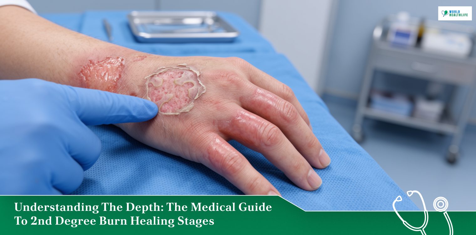
The human skin is a remarkably resilient shield that acts as a critical barrier against environmental trauma, fluid loss, and bacterial infection.
When a thermal, chemical, or electrical accident occurs, this biological shield sustains structural damage that requires a highly coordinated and cellular recovery effort.
Among the various classifications of skin trauma, a partial-thickness injury presents some of the most complex clinical challenges.
When you seek precise answers regarding the 2nd degree burn healing stages, it is vital to recognize that recovery is not a single, uniform event.
Instead, the body initiates a highly regulated, multi-layered physiological pipeline to clear away dead tissue, rebuild the dermal matrix, and restore the skin’s original tensile strength.
Navigating this recovery process requires moving past basic first-aid checklists.
Reclaiming your skin health requires an intimate, realistic look at the distinct biological phases of tissue repair.
Thus, the following guide will focus on these phases while delving deep into the explicit quadrant signs that track your daily progress.
Finally, it will evaluate the crucial medical boundaries needed to prevent permanent complications.
Defining The Trauma: What Really Is A Partial-Thickness Injury
Medical professionals classify a second-degree burn. This is clinically referred to as a partial-thickness burn by its depth of tissue destruction.
Unlike a superficial injury that only damages the top layer of skin, this specific trauma cuts deeper into the underlying anatomy.
A second-degree burn completely destroys the epidermis while penetrating straight into the dermis. This layer houses sweat glands, hair follicles, blood vessels, and nerve endings.
Since the structural integrity of the dermal layer is compromised, blood capillaries leak fluid into the surrounding tissue.
As a result, this creates the signature fluid-filled blisters.
Simultaneously, the raw exposure of delicate nerve endings triggers an intense, burning pain. This pain makes partial-thickness injuries uniquely distressing.
Clinicians further divide these burns into:
- superficial partial-thickness burns, which damage only the upper dermis
- deep partial-thickness burns, which destroy deeper dermal layers
This completely dictates the overall timeline across the 2nd degree burn healing stages.
Categorizing The Spectrum: Different Classifications Of Burns
In order to understand how a partial-thickness injury heals, it helps to place it within the wider context of thermal skin damage.
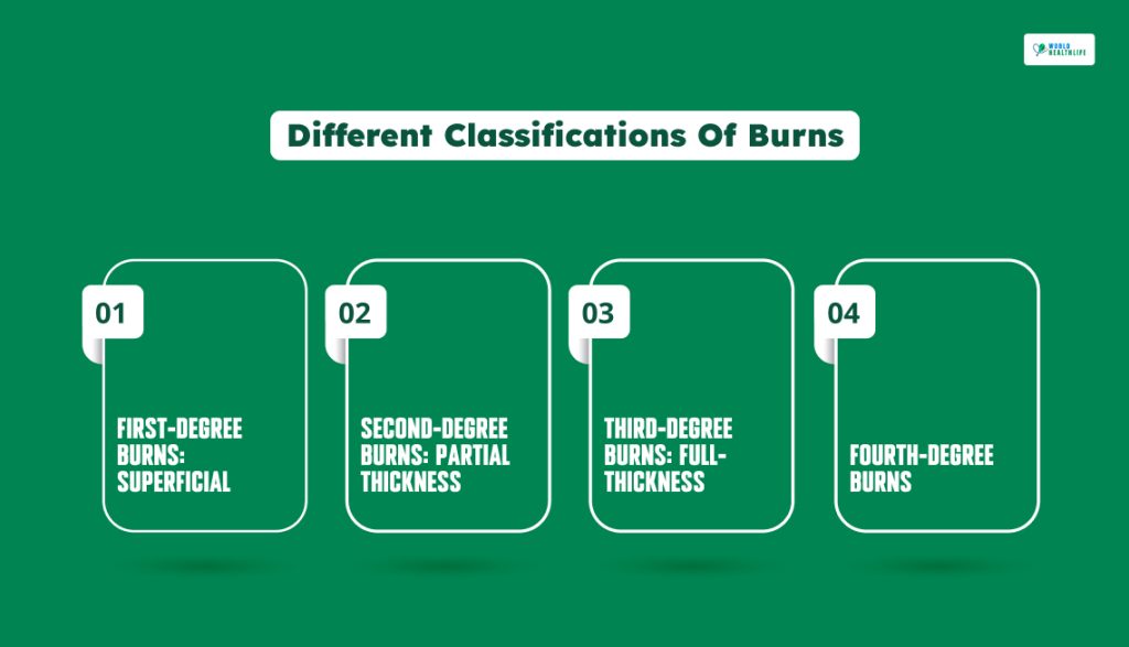
The medical community relies on four primary structural classifications to evaluate skin trauma.
First-Degree Burns: Superficial
The initial first-degree burns impact the epidermis alone. The skin looks pink or red, feels dry, and temporarily turns white when pressed. This white area is “blanched.”
There are no blisters on the surface, typically healing cleanly within three to six days without leaving a permanent scar.
Second-Degree Burns: Partial Thickness
This classification destroys the entire outer layer and penetrates straight into the structural dermis.
The tissue appears moist, bright red, or mottled white, and it features prominent fluid-filled blister formation.
Because nerve endings are exposed, it is paired with exquisite pain sensitivity.
Third-Degree Burns: Full-Thickness
This severe trauma completely destroys both the epidermis and the entire depth of the dermis. This extends down into the subcutaneous fat layer.
The skin looks leathery, waxy, white, or charred black.
Since the underlying nerve infrastructure is entirely incinerated, the wound itself is completely numb to the touch.
Fourth-Degree Burns
This is the most catastrophic tier of thermal injury.
The destruction extends past the skin layers to incinerate underlying muscle groups, deep tendons, ligaments, and bone structures.
An immediate specialized surgical intervention is required in this phase.
Chronological Breakdown Of The 2nd Degree Burn Healing Stages
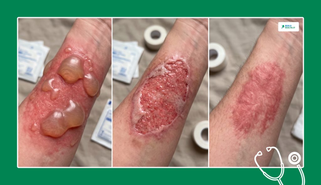
The biological reconstruction of a partial-thickness injury follows a strict and overlapping cellular timeline.
For the skin to close cleanly without a secondary breakdown, the body must systematically progress through three distinctive operational phases.
Phase 1: The Inflammation Or Reactive Stage
The moment a thermal accident occurs, the body initiates an immediate survival response to stabilize the tissue.
This reactive phase serves as an automated protective barrier before actual tissue rebuilding can begin.
What Happens
First, local blood vessels constrict to stop fluid loss. This is followed rapidly by a widening of the vessels, or vasodilation.
This allows plasma, signaling proteins, and specialized white blood cells such as neutrophils and macrophages to flood the wound bed.
These cells act as an internal cleanup crew, systematically swallowing up scorched tissue fragments.
Furthermore, it goes on to neutralize foreign bacteria and establish a clean foundation for new skin growth.
Timeline
This initial reactive phase begins within seconds of the injury and remains active for roughly the first 1 to 5 days of the recovery process.
Signs
The wound bed looks highly inflamed, swollen, and bright red.
You will experience intense localized pain, a hot radiating sensation around the tissue, and active fluid leakage from ruptured blisters as plasma continuously washes the area.
Phase 2: The Proliferation Or Reparative Signs
Once the wound bed is successfully cleared of debris, the body transitions from containment to structural rebuilding.
This phase is characterized by rapid cellular division as new tissue layers fill in the open gap.
What Happens
Specialized cells called fibroblasts migrate to the injury site and begin pumping out a fresh scaffolding of collagen fibers.
Concurrently, the body initiates angiogenesis, which is the rapid creation of tiny new blood capillaries. This is done to deliver vital oxygen and nutrients to the growing tissue.
This creates a moist, pink, bumpy tissue layer known as granulation tissue.
Over this fresh base, epithelial cells from the wound edges and surviving hair follicles multiply and crawl across the gap to form a new outer skin layer.
Timeline
This intensive rebuilding phase overlaps with inflammation, typically around day 4 and lasts up to day 21. This is for superficial partial-thickness injuries.
Signs
The continuous fluid weeping drops off significantly. The wound bed begins to fill in with moist, healthy pink or deep red granulation tissue.
You will notice the edges of the burn tightening inward as a thin, translucent layer of fresh pink skin slowly sheets over the open wound.
Phase 3: The Maturation Or Remodeling Stage
With the outer skin layer successfully sealed, the body shifts its focus from rapid building to long-term structural reinforcement.
This final stage is the longest part of the recovery process, quietly adjusting the deep tissue layout beneath the surface.
What Happens
Enzymes systematically break down the disorganized, fragile collagen fibers laid down during proliferation.
Instead, highly organized, dense collagen bundles replace it, aligning with the skin’s natural tension lines.
This process dramatically increases the skin’s tensile strength, though the remodeled tissue will permanently max out at about 80% of its original pre-burn elasticity.
Timeline
This final phase is an incredibly long, slow process.
It begins around week 3 and can continue for up to 1 to 2 full years depending on the original depth of the dermal damage.
Signs
The intense redness of the fresh skin slowly fades away, changing to a pale pink, white, or hyperpigmented skin tone.
This may match or deepen your natural skin color. The fresh tissue may feel abnormally stiff, tight, or thick.
Additionally, it is frequently accompanied by a persistent, deep itch as local nerve fibers slowly repair themselves underneath the surface.
Vital Pre-Care Boundaries: Essential Precautions For Safe Wound Closure
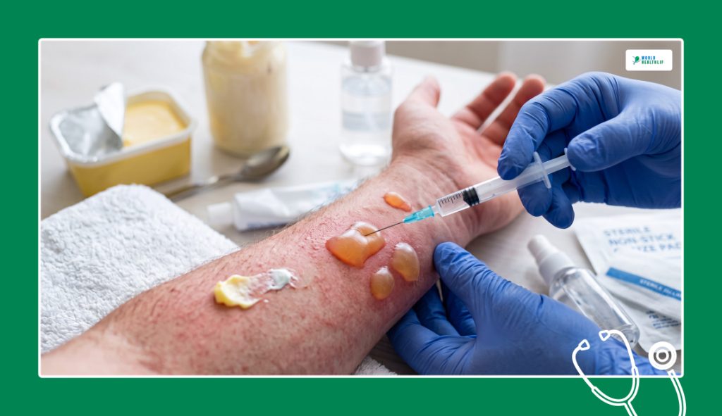
How you treat a partial-thickness injury during its initial reactive phase directly shapes your trajectory across the later 2nd degree burn healing stages.
Therefore, implementing the wrong first-aid choices can accidentally turn a clean, superficial wound into a deep, infected surgical crisis.
Never Pop Fluid-Filled Blisters
The blister bubbles that form on a partial-thickness burn serve as a sterile and natural bandage created by your own immune system.
Moreover, the blister fluid contains vital growth factors that speed up early cellular repair.
Popping these bubbles strips away this sterile protection.
Accordingly, it leaves a raw and open doorway for aggressive surface bacteria to enter and trigger a deep tissue infection.
Stop Using Folk Remedies
Never apply butter, heavy cooking oils, mayonnaise, or toothpaste to a fresh burn.
These thick substances create an artificial seal over the wound, trapping the thermal heat deep inside your tissue layers. This causes the burn to continue cooking downward into the dermis.
Furthermore, these unsterile ingredients introduce harmful bacteria straight into compromised tissue. Thus, this increases the likelihood of permanent tissue scarring.
Comprehensive Recovery Matrix
To safely manage a partial-thickness injury across the long-term 2nd degree burn healing stages, your daily care routine must shift on the basis of the specific biological needs of each phase.
Phase-Specific Clinical Guidelines
| Healing Phase | Focus Metric | Core Tactical Care Protocol | Critical Complication Warning Signs |
| Inflammation | Infection prevention and thermal stabilization. | Cool the tissue with clean, room-temperature water; apply sterile, non-stick dressings. | Presence of foul-smelling drainage, streaks of red tracking up the limb, or a sudden fever. |
| Proliferation | Moisture balance and cellular crawling support. | Apply prescribed antimicrobial ointments (like silver sulfadiazine); protect fresh granulation layers. | Sudden darkening or death of the pink tissue, or a complete halt in edge tightening. |
| Maturation | Tissue flexibility and structural scar mitigation. | Gently massage with pure, unscented emollient creams; protect the area with SPF 50 sunblock. | Development of thick, raised keloid scars, or severe joint contractures that restrict movement. |
Knowing When To Pivot To Emergency Clinical Care
You can successfully manage many minor partial-thickness injuries at home by carefully tracking the 2nd-degree burn healing stages.
However, specific clinical red flags require immediate emergency room intervention.
A second-degree burn may cover an area larger than three inches in diameter or affect highly sensitive anatomical zones. These zones include the face, major joints, hands, feet or the groin.
In that case, it is necessary that you seek professional burn center care immediately.
Furthermore, if your pain levels suddenly spike days after the accident, or the surrounding skin becomes intensely hot, swollen, and tracks red lines toward your heart, your wound has likely sustained a secondary bacterial infection.
This shift requires immediate prescription antibiotic therapy to protect your systemic health and preserve your long-term skin integrity.
Disclaimer: The information provided in this article is for general informational purposes only. It does not, and is not intended to, constitute medical or health advice. Please consult a qualified healthcare professional for medical guidance or expert health assistance.
References
- Cleveland Clinic. (2025). Second-Degree burn. Cleveland Clinic.
- Releford, B. (2023). The Stages of Second-Degree Burn Healing: What to expect. The Wound Pros.
- Gupta, S., Chittoria, R. K., Chavan, V., Aggarwal, A., Reddy, L. C., Mohan, P. B., Koliyath, S., & Pathan, I. (2021). Role of Burn Blister Fluid in Wound Healing. Journal of Cutaneous and Aesthetic Surgery, 14(3), 370. National Library of Medicine.


