The human brain relies on a delicate balance of minerals in order to transmit signals in a smooth manner across its neural networks.
Conversely, with the failure of this mineral balance, calcium starts to silently pool inside the brain’s deep motor control centers.
Additionally, over several decades, these microscopic mineral deposits harden, essentially turning vital brain tissue into stone.
This underlying process defines the reality of Fahr’s disease, an ultra-rare and progressive neurological disorder. Widespread and symmetrical brain calcification characterizes the condition.
First mapped out historically in the early 20th century, this genetic condition targets the basal ganglia.
This portion is the primary control station of the brain, which is responsible for controlling movement, emotional regulation, and cognitive processing.
One thing that you should know is that Fahr’s disease is not an acute illness that develops overnight. Instead, it acts as a slow-moving degenerative process that slowly cuts off the communication lines between the brain and the body.
To understand this complex condition, you would require looking past the generic definitions of aging or standard memory loss. Navigating life with this diagnosis takes a raw and realistic look at certain things.
Thus, the following guide, keeping exactly that in mind, will cover its genetic origins, its overlapping movement and psychiatric symptoms, and what it actually takes to manage the daily physical fallout.
Defining The Basic Mechanism: Genetic Faults Vs. Secondary Calcification
Medical professionals use the term Fahr’s disease to describe a primary, familial genetic condition.
Do not confuse it with Fahr’s syndrome, which presents with similar brain calcification but stems from secondary metabolic disorders like hypoparathyroidism.
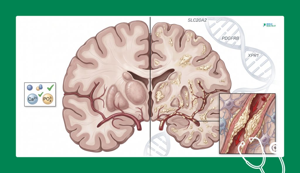
In its primary genetic form, the body’s overall calcium and phosphate levels in the blood appear completely normal.
The issue is not an excess of calcium in the body. Instead, it is a localized failure of the blood vessels of the brain to keep minerals where they belong.
The illness is primarily an autosomal dominant genetic disorder. If a parent carries the mutated gene, their children face a literal fifty-fifty chance of inheriting the condition.
Researchers have traced the root cause of Fahr’s disease to mutations in a few specific genes, most notably SLC20A2, PDGFRB, and XPR1.
For instance, the SLC20A2 gene provides the instructions to build a specific inorganic phosphate transporter protein in the brain.
A flaw in this gene causes phosphate levels to build up inside the walls of the cerebral blood vessels.
Consequently, this localized chemical imbalance acts like a magnet, drawing calcium out of the bloodstream and binding it directly into the surrounding tissue.
Over time, these mineral deposits restrict blood flow and choke out nearby neurons, causing the globus pallidus, caudate nucleus, and putamen to slowly degenerate.
The Four Diagnostic Strands of Brain Calcification
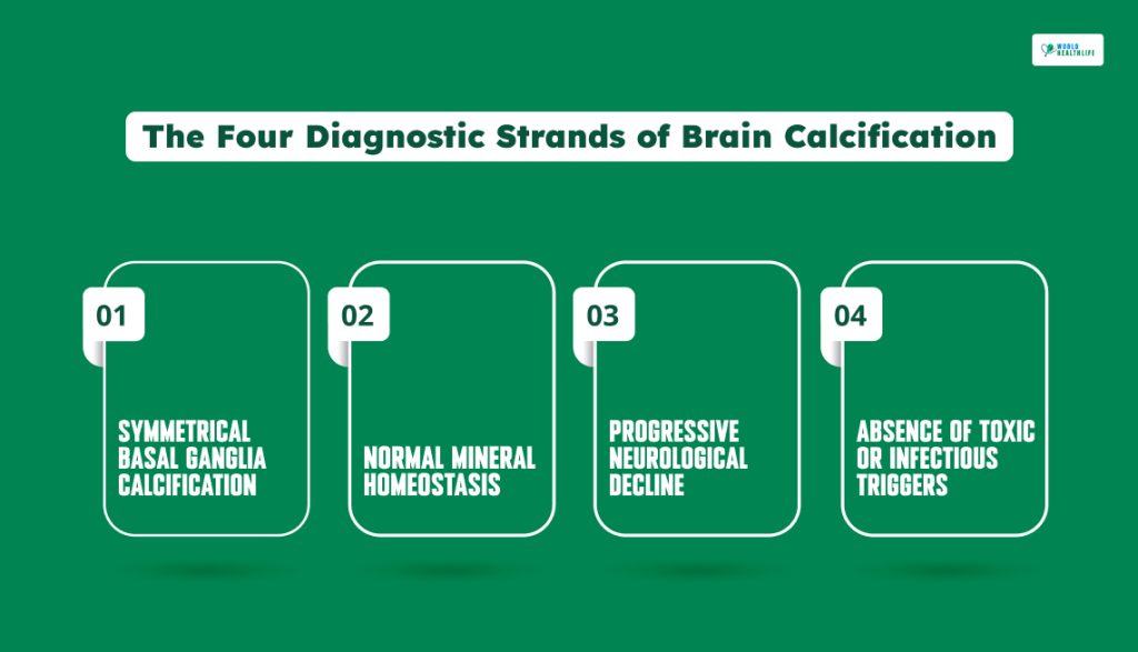
Confirming a case requires matching targeted neuroimaging with a comprehensive blood work panel to isolate the primary genetic variant from secondary lookalikes.
Clinicians look for four specific pillars to lock in a diagnosis of Fahr’s disease.
Symmetrical Basal Ganglia Calcification
High-resolution computed tomography (CT) scans must show dense and bone-like mineral blocks distributed evenly across both sides of the basal ganglia.
Normal Mineral Homeostasis
Laboratory blood tests must show completely normal ranges for serum calcium, phosphorus, parathyroid hormone (PTH), and alkaline phosphatase.
Progressive Neurological Decline
The patient must present documented, advancing motor or cognitive deficits that match the areas of brain tissue loss seen on the scans.
Absence Of Toxic Or Infectious Triggers
Clinical teams must explicitly rule out any history of brain radiation, heavy metal poisoning, mitochondrial illnesses, or chronic intrauterine infections.
Mapping The Split Clinical Presentation: Movement Vs. Mind
The symptoms of Fahr’s disease typically lie dormant until early to mid-adulthood, with most patients noticing the first warning signs between the ages of 30 and 50.
Because the basal ganglia control both physical motion and emotional processing loops, the disease fractures into two distinct clinical pathways.
| Clinical Dimension | Primary Symptoms & Presentation | Real-World Functional Impact |
| Extrapyramidal Movement | Progressive parkinsonism, including severe muscle rigidity, a shuffling gait, and resting tremors. | Patients struggle with fine motor skills, lose independent balance, and experience frequent, unprovoked falls. |
| Hyperkinetic Elements | Involuntary, dancing limb movements (chorea) and painful, twisting muscle contractions (dystonia). | Continuous spasms twist the limbs or neck into unnatural shapes, causing chronic joint pain and exhaustion. |
| Psychiatric & Behavioral | Severe, treatment-resistant depression, sudden mood swings, apathy, and intense paranoia. | Personality shifts strain family relationships and can lead to unprovoked outbursts or social withdrawal. |
| Cognitive & Executive | Accelerated memory loss, a drop in processing speed, and poor concentration. | Progresses into a subcortical dementia where patients lose the ability to plan, make decisions, or live independently. |
Visualizing The Damage On Neuroimaging
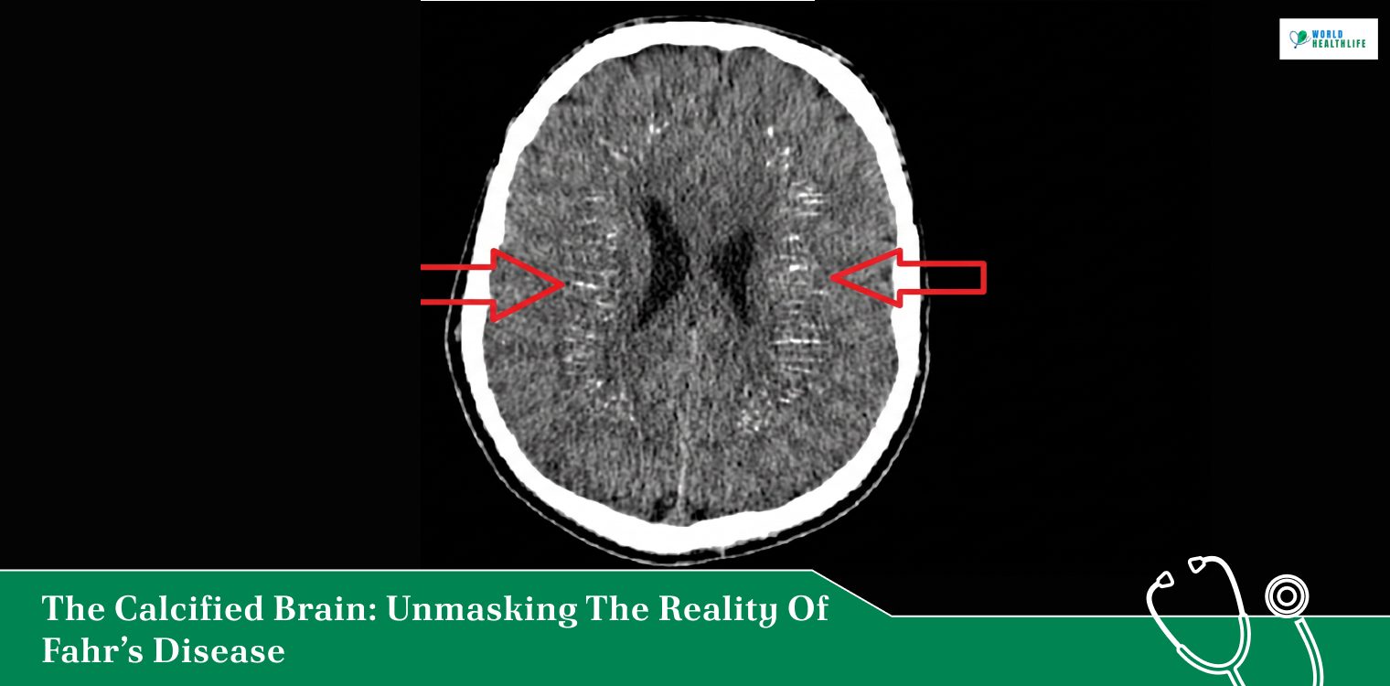
The single most important diagnostic tool for evaluating Fahr’s disease is a computed tomography (CT) scan.
Although magnetic resonance imaging (MRI) is excellent for viewing soft tissue, CT scans are vastly superior at spotting hard mineral deposits.
On a typical head CT, the calcified zones light up in a brilliant, stark white, matching the exact density of the surrounding skull bones.
These bright white patches outline a footprint of destruction. In the early stages, the calcification localized strictly to the globus pallidus.
But as Fahr’s disease advances, the white patches spread outwards like a map, consuming the thalamus, the cerebral cortex, and the internal capsule.
The denser and wider these white blocks appear on the scan, the more severe the patient’s physical stiffness and cognitive deficits tend to be.
Navigating The Care And Treatment Pipeline
Unfortunately, there is currently no way to dissolve calcium deposits inside the brain or reverse the structural damage.
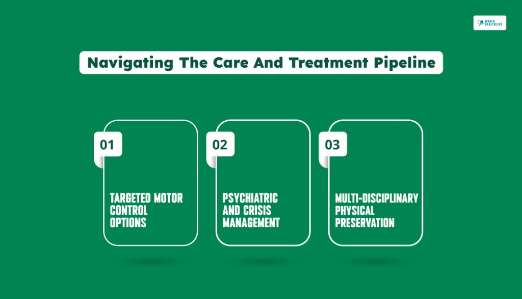
Since a universal cure does not exist, medical teams focus entirely on managing individual symptoms. Moreover, they tend to put their focus on preserving daily mobility and soothing psychiatric distress.
Targeted Motor Control Options
To manage the stiff, Parkinsonian movement issues, neurologists often prescribe levodopa or dopamine agonists.
However, Fahr’s disease destroys the underlying brain receptors rather than just dropping dopamine production.
As a result, these medications often provide only partial, temporary relief. For patients dealing with painful, localized muscle twisting, targeted botulinum toxin (Botox) injections can help relax rigid muscle groups and reduce chronic pain.
Psychiatric And Crisis Management
Managing the chemical shifts in the brain requires a careful approach to mental health.
Standard antidepressants (SSRIs) are routinely deployed to balance sudden mood drops and anxiety.
However, if a patient develops intense paranoia or psychosis, clinicians must use anti-psychotic medications with extreme caution.
Many typical antipsychotics can severely worsen the patient’s existing physical stiffness and tremors, requiring specialized, atypical options instead.
Multi-Disciplinary Physical Preservation
Because the disease damages swallowing and speech pathways over time, early speech therapy is essential to prevent choking risks.
Concurrently, intensive physical and occupational therapy regimens help patients with a variety of activities including:
- stretch stiff limbs
- maintain independent balance
- learn to use adaptive mobility aids before coordination drops sharply.
Shifting Expectations For Long-Term Adaptation
Fundamentally, for any family, getting a diagnosis of Fahr’s disease alters their overall trajectory.
The progression is slow but unyielding, requiring ongoing home modifications, specialized wheelchairs, and intensive physical care as the years roll by.
Since the condition cannot be cured by a standard medical intervention, the core focus of care requires something else.
Instead, it must shift toward maximizing daily comfort, minimizing physical pain, and preserving communication lines.
Ultimately, it can be recommended to combine precise CT imaging diagnostics with early, aggressive symptom management and genetic family counseling.
Clinical teams can help families face this complex calcification process with absolute dignity and long-term resilience.
Disclaimer: The information provided in this article is for general informational purposes only. It does not, and is not intended to, constitute medical or health advice. Please consult a qualified healthcare professional for medical guidance or expert health assistance.
References
- Damasceno Zanuto Guerra, A. C., Pessoa, C. H., Marquezine, G. F., & Alvares Delfino, V. D. (2021). Hypoparathyroidism and Fahr’s syndrome: Case series. Jornal Brasileiro de Nefrologia, 44(4), 592. National Library of Medicine.
- Manyam, B. V. (2005). What is and what is not ‘Fahr’s disease’. Parkinsonism & Related Disorders, 11(2), 73-80. ScienceDirect.