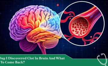
Structural symmetry fundamentally builds the human brain.
Although each hemisphere handles different tasks, both sides grow at the exact same pace during fetal development. As a result, there is a balanced and evenly proportional organ.
But sometimes, a rare genetic glitch disrupts this blueprint before birth. In hemimegalencephaly, one half of the brain grows massively out of control.
Consequently, it starts becoming abnormally large and structurally deformed, while the other half remains completely normal. Experts consider this an ultra-rare and congenital brain malformation that bypasses traditional neurological rules.
The condition is not some disease a child catches or a slow-moving degenerative illness. Instead, it is a structural overload present entirely from conception.
The enlarged side of the brain features scrambled neural wiring and abnormal cells that constantly misfire. This mismatch triggers a wave of early-onset seizures, developmental delays, and motor challenges.
Managing this condition requires moving past basic textbook definitions. Families and medical teams must look closely at its genetic triggers, its distinct imaging signs, and the surgical paths needed to help a child find balance.
The Structural Defect In Hemimegalencephaly: An Overgrowth Of One Hemisphere

Before we dive deep into the topic, first let us talk about what exactly the condition is.
Medical professionals classify hemimegalencephaly as a disorder of cellular proliferation and migration.
During early embryonic development, the stem cells that build the cerebral cortex must divide evenly.
In this condition, a localized genetic mutation causes the cells on one side to multiply excessively. At the same time, these cells fail to migrate to their proper positions.
As a result, this leaves the enlarged hemisphere with a deeply scarred, thickened and disorganized cortex.
The structural impact goes far beyond a simple size difference, where the enlarged side often displays two things.
- Pachygyria: Abnormally thick and flat folds
- Polymicrogyria: An excessive number of tiny and malformed folds
Deep within the tissue, the normal boundaries between gray matter and white matter blur entirely.
Furthermore, the cells themselves become warped, where neurologists frequently find balloon cells. These are bizarre and giant cells that share characteristics of both neurons and glial cells.
Because this overgrown tissue is structurally unstable, it acts like a massive electrical storm. This constantly sends chaotic signals across the midline while disrupting the function of the healthy hemisphere.
The Genetic Blueprint: Somatic Mosaicism And The mTOR Pathway
Hemimegalencephaly stands out because parents rarely pass it down, and it almost never runs in families.
Instead, a post-zygotic mutation drives the condition. Additionally, a genetic error happens randomly after the egg is fertilized, during the early stages of cell division in the womb. Scientists call this biological phenomenon somatic mosaicism.
This mutation targets the mTOR pathway, which is the mammalian target of rapamycin
Only the cells descending from that mutated lineage will grow abnormally. This is why the defect remains strictly confined to one half of the brain.
The condition can occur entirely on its own as an isolated anomaly. Besides that, it can also present as part of a wider neurocutaneous syndrome, such as Epidermal Nevus Syndrome, Hypomelanosis of Ito, or Proteus syndrome.
The Four Major Diagnostic Indicators On Brain MRIs

Confirming a case requires high-resolution neuroimaging, which is specifically a T2-weighted magnetic resonance imaging (MRI) scan.
Radiologists look for a classic cluster of signs to separate hemimegalencephaly from other overgrowth disorders.
Asymmetric Cranial and Ventricular Overgrowth
The skull itself is often asymmetric, visibly bulging on the side of the defect.
On the MRI screen, the lateral ventricle within the enlarged hemisphere is stretched out and dilated. Oftentimes, this takes on a straight, pointed shape known as a “straight ventricular horn.”
Scrambled Cortical Architecture
The normal, clean folds of the brain are replaced by thick, disorganized tissue patches.
The white matter looks abnormally bright on the scan, showing signs of premature or flawed myelination. On the other hand, the normal boundary line separating gray and white matter disappears completely.
Displacement of the Posterior Fossa
The sheer mass of the overgrown hemisphere physically pushes against the surrounding structures.
This crowding forces the brain’s midline cleft to curve into a distinct, C-shaped arc, pushing the brainstem and the cerebellum out of place toward the healthy side.
Contralateral Structural Integrity
A vital diagnostic marker is that the opposite hemisphere remains completely normal in size and structure.
This stark contrast between a healthy half and an overgrown half is the absolute signature of hemimegalencephaly. Consequently, this allows clinical teams to rule out global megalencephaly or diffuse tumors.
Spotting The Signs: Clinical Presentation And Symptoms
Since one half of the brain is constantly misfiring, the condition triggers an aggressive mix of physical and neurological symptoms very early in life.
| Clinical Area | Typical Presentation and Evolution | Long-Term Development |
| Seizure Activity | Starts in early infancy, often as infantile spasms or continuous focal seizures. | Highly resistant to standard anti-epileptic drugs; can happen dozens of times a day. |
| Motor Function | Weakness or partial paralysis on the opposite side of the body (hemiparesis). | The child struggles with core balance, displays stiff muscles, and experiences severe motor delays. |
| Cognitive Profiles | The constant electrical storms disrupt early learning milestones. | Leads to intellectual disabilities and developmental arrest if seizures aren’t controlled. |
| Visual Pathways | Involuntary, rhythmic eye jerking (nystagmus) and crossed eyes (strabismus). | Hemianopia develops, leaving the child blind in the visual field opposite the deformed hemisphere. |
Navigating The Treatment Pipeline: From Drugs To Disconnection

There is no way to chemically shrink the enlarged tissue or fix the broken genetic pathway. Since the standard anti-seizure medications almost completely fail to control the episodes, medical teams need to pivot to aggressive interventions.
Accordingly, this helps to stop the constant electrical storms while protecting the healthy side of the brain.
Medical Management Limits
Neurologists will initially try combinations of aggressive anti-epileptic drugs, or a medically supervised ketogenic diet. This allows the brain’s chemistry to change.
In cases that have a direct relation to the mTOR pathway, researchers make active efforts in exploring targeted therapies.
Therapies such as mTOR inhibitors (everolimus and other types) are used to see if they can slow down abnormal cell growth.
However, for most children, medications are just a temporary band-aid.
Anatomical Hemispherectomy
When seizures remain uncontrollable while threatening to ruin the healthy half of the brain, a radical surgical procedure known as anatomical hemispherectomy becomes the primary option.
In this operation, neurosurgeons completely remove the physical tissue of the diseased, overgrown hemisphere.
Functional Hemispherectomy Disconnection
Alternatively, surgeons may choose a functional hemispherectomy.
Instead of taking out the entire half of the brain, they leave the physical tissue in place.
However, all the white-matter tracts connecting it to the rest of the body should be surgically sliced through.
Accordingly, this completely disconnects the electrical storms from the healthy side, stopping the seizures from spreading.
The Path Forward: Embracing Neuroplasticity
Hearing a diagnosis of hemimegalencephaly can completely bring a total change to any family’s life.
The medical choices are heavy, often forcing parents to choose a radical brain surgery that permanently sacrifices fine motor control on one side of the body.
Despite that, the ability of the brain to adapt after a hemispherectomy is a marvel of biological resilience.
Since the surgery is performed early in life, a child’s remaining healthy hemisphere displays incredible neuroplasticity. Over time, this single, healthy half can rewire itself, taking over critical speech, walking, and cognitive tasks.
By choosing the right surgical timing and backing it up with intensive physical therapy, medical teams can stop the electrical storms.
As a result, these children are given a real chance to rebuild their lives with a single, balanced half.
Disclaimer: The information provided in this article is for general informational purposes only. It does not, and is not intended to, constitute medical or health advice. Please consult a qualified healthcare professional for medical guidance or expert health assistance.