
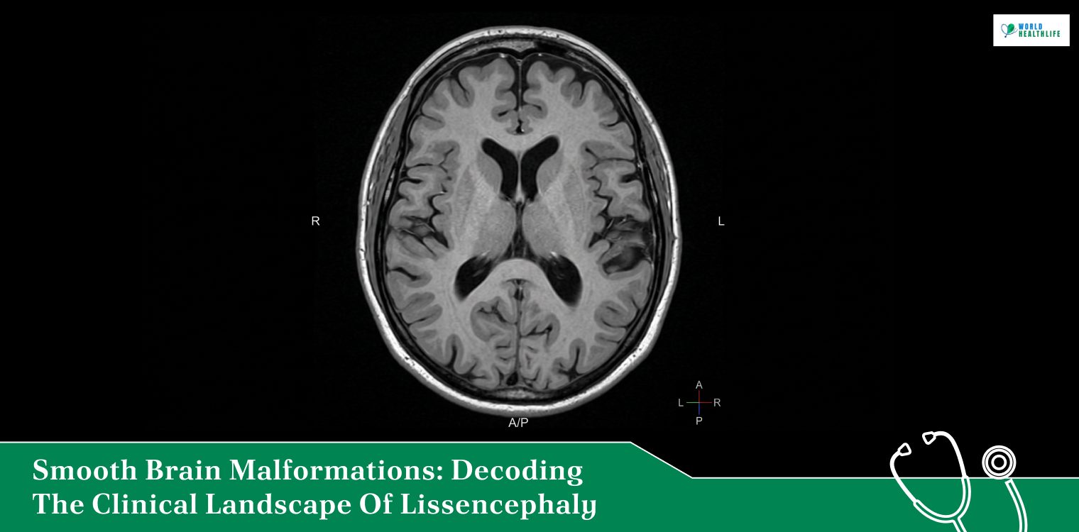
The human brain is a marvel of complex geography, where its deeply folded surface is covered in intricate ridges known as gyri and grooves called sulci. These ridges are not just for show.
Instead, this unique setup allows nature to pack up a massive amount of computing power into the tight space of the skull.
This folding process is absolutely vital for the way we think, move and process the world.
However, sometimes a rare genetic breakdown halts this structural blueprint mid-construction. This condition is known as lissencephaly, which literally translates from Greek as “smooth brain.”
Instead of developing a highly folded surface, the brain remains flat. It either lacks ridges entirely or possesses only a few thick and poorly formed grooves.
Since a smooth cortex lacks the necessary organizational layers to route signals correctly, children born with this condition face certain extreme issues:
- Severe epilepsy
- Profound developmental delays
- Complex motor challenges
Navigating life with this diagnosis requires a raw and realistic look at its general triggers as well as treatment realities.
The Developmental Error: A Breakdown In Neuronal Migration
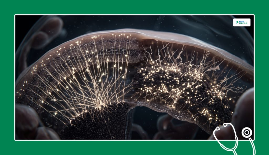
In order to understand the architecture of a smooth brain, you have to look at the way a child’s nervous system builds itself during the first two trimesters of pregnancy.
The Cellular Journey
Between the 12th and 24th weeks of gestation, a massive and highly coordinated cellular migration takes place.
Billions of newborn nerve cells, or neurons, are generated deep inside the center of the embryonic brain, near the fluid-filled ventricles.
Once created, these young neurons are required to travel outward to the surface of the brain to form the six distinct layers of the cerebral cortex.
They do this by climbing along specialized guidelines called radial glial fibers, moving outward like rock climbers scaling a wall.
The Premature Stop
In lissencephaly, this highly orchestrated migration grinds to a sudden halt. The young neurons begin their journey normally. However, they lose their way or stop climbing long before they can reach their destination.
Instead of forming six highly organized, thin layers of grey matter, the cells get stuck in transit. Moreover, they bunch together into a thick and disorganized four-layered cortex.
Since the neurons never reach the outer surface to expand the brain’s surface area, the outer cortex remains flat and smooth.
This underlying migration failure is classified as a classic disorder of neuronal migration. In addition, its severity depends entirely on when and where the cellular journey was disrupted.
Genetic Driver Configurations: The LIS1 And DCX Pathways
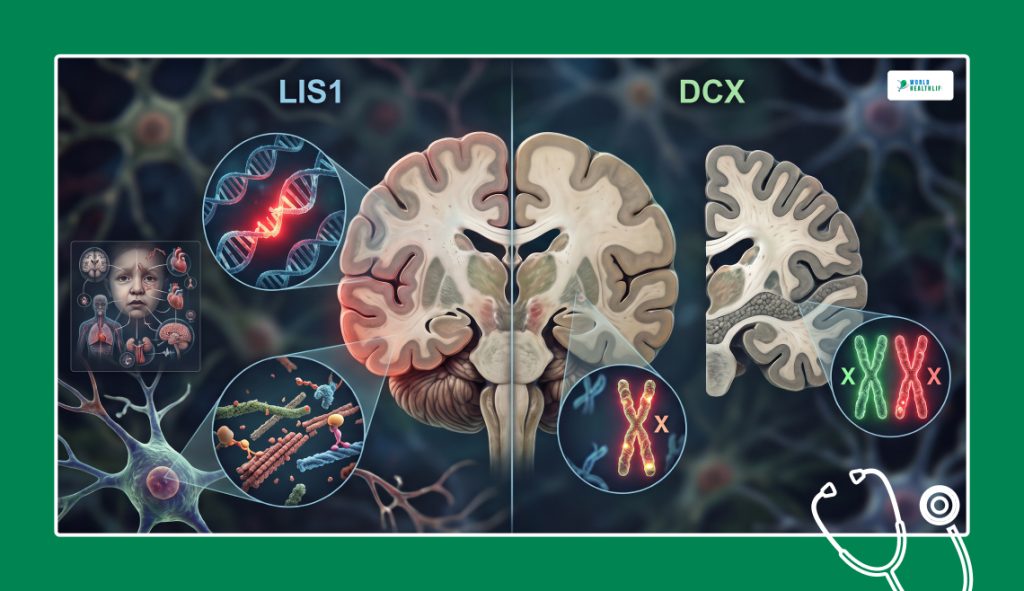
Environmental factors such as intrauterine or a sudden loss of fetal blood supply can occasionally disrupt cellular movement. However, these specific genetic mutations drive the vast majority of cases.
The LIS1 Gene Mutation: Chromosome 17
Mutations or deletions in the LIS1 gene disrupt the production of a protein. Moreover, this protein is absolutely vital for maintaining the structural integrity of the cellular motor network inside migrating neurons.
A flawed LIS1 gene typically causes severe diffuse smoothness that is most pronounced at the back of the brain.
The complete deletion of a large section of chromosome 17 containing the gene causes the child to develop a specific condition.
Known as Miller-Dieker syndrome, the condition couples the smooth brain malformation with distinct facial traits and severe systemic complications.
The DCX Gene Mutation: The X-Linked Pathway
The DCX gene provides the instructions to build doublecortin, a protein responsible for the stabilization of the internal scaffolding of a neuron as it moves.
Because the DCX gene is located on the X chromosome, its clinical impact depends heavily on the biological sex of the child.
In Case Of Males
In biological males, a mutated DCX gene causes severe and widespread lissencephaly. The reason behind this is that boys carry only one X chromosome.
Typically, their cases feature an anterior-dominant pattern where the front of the brain is the smoothest.
In Case Of Females
Conversely, in females, they carry a second, healthy X chromosome to offset the mutation.
Consequently, this only partially disrupts cellular migration. This creates a fascinating structural anomaly known as subcortical band heterotopia, or “double cortex syndrome.”
Here, a smooth band of trapped grey matter sits directly beneath a normally folded outer layer.
Structural Severity Scales On Neuroimaging
The confirmation of a diagnosis requires high-resolution neuroimaging. That is, specifically, a magnetic resonance imaging (MRI) scan.
Radiologists do not just look for a completely smooth surface. Instead, they use specific structural criteria to grade the severity of the malformation. This is based on the degree of cortical thickening and the presence of residual grooves.
| Severity Grade | Radiological Classification | Cortical Surface Appearance | Structural White/Grey Matter Boundaries |
| Grade 1 | Complete Agyria | Totally smooth cortex across the entire brain; zero sulci or gyri visible. | Widespread, severe cortical thickening; the normal six-layer gray matter boundary is completely absent. |
| Grade 2 | Regional Agyria / Pachygyria | Complete smoothness at the back of the brain, transitioning to a few thick, flat folds at the front. | Thick, disorganized gray matter bands with a significant reduction in underlying white matter tracks. |
| Grade 3 | Diffuse Pachygyria | No completely smooth zones, but the entire brain surface is covered in fewer, unusually thick, wide ridges. | Broad, simplified cortical layers with mild dilation of the lateral ventricles. |
| Grade 4 | Subcortical Band Heterotopia | A fully folded, normal outer cortex sitting directly above a distinct, smooth band of trapped gray matter cells. | Unmistakable “double cortex” appearance separated by a thin layer of healthy white matter. |
Spotting The Complications: Clinical Presentation And Symptoms
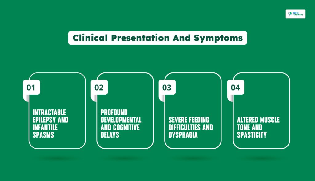
Since a smooth cortex cannot route electrical signals smoothly, the condition triggers an intense and interconnected web of neurological and physical symptoms.
These start to appear very early in infancy.
Intractable Epilepsy And Infantile Spasms
The single most dominant and difficult aspect of lissencephaly is the development of severe, drug-resistant epilepsy.
Over 90% of children with this condition develop seizures before their first birthday.
These frequently begin as infantile spasms, which are sudden, violent clusters of body flexion.
Additionally, these can happen dozens of times a day, disrupting early brain activity and contributing to developmental arrest.
As the child grows, these spasms often evolve into complex, multi-focal generalized seizures that standard medications struggle to control.
Profound Developmental And Cognitive Delays
The brain struggles to process and store complex information because the completely disorganized structural layers of the cortex disrupt its function.
Children typically experience severe intellectual disabilities. Many do not achieve standard milestones, requiring lifelong, around-the-clock care.
These include:
- rolling over
- sitting up independently
- tracking objects visually
- developing verbal communication
Severe Feeding Difficulties And Dysphagia
The neurological structural deficits directly impact the cranial nerves responsible for managing the mouth and throat muscles.
Infants frequently suffer from severe dysphagia (difficulty swallowing) and poor sucking reflexes.
As a result, feeding becomes a significant hazard, as food or liquid can easily slip into the airway.
This silent choking, known as aspiration, leads to a high risk of recurring aspiration pneumonia.
Thus, it makes surgical feeding tubes (G-tubes) a common necessity to protect the child’s lungs and maintain steady nutrition.
Altered Muscle Tone And Spasticity
The motor pathways coming down from the smooth cortex send erratic, uncoordinated signals to the body.
Infants often present with severe hypotonia, which is a floppy, weak core and neck in early life.
As the nervous system matures, this floppiness typically stiffens into severe spasticity. Rigid, tight limbs, restricted joint movement, and involuntary muscle contractions result.
Navigating the Care And Treatment Pipeline
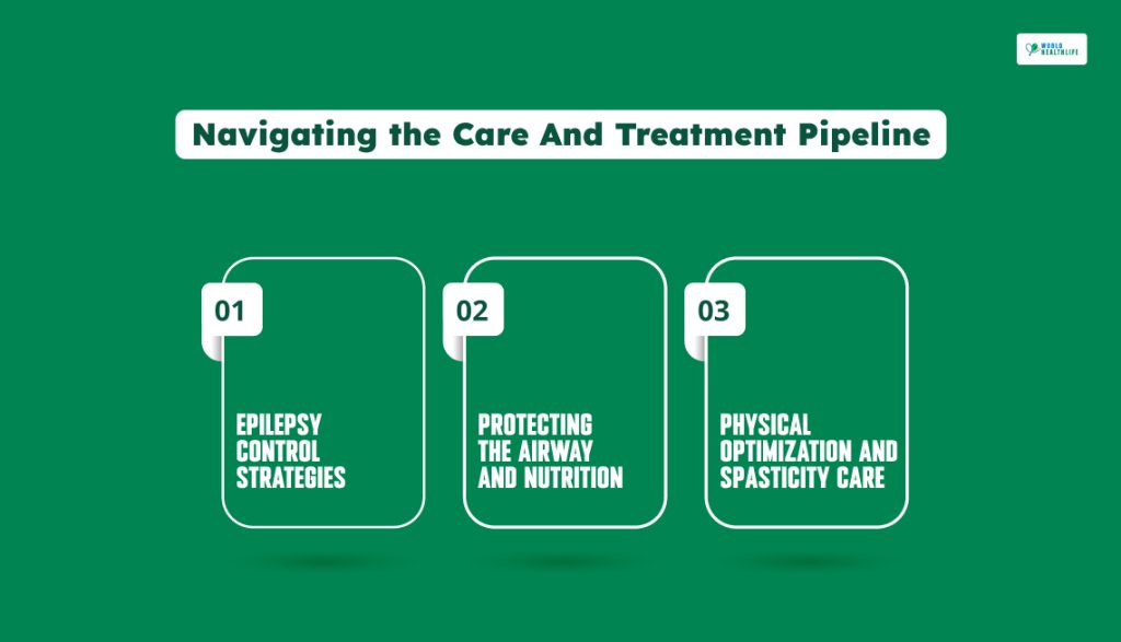
There is currently no way to surgically induce folding on a smooth cortex, nor is there a gene therapy capable of restarting neural migration once a child is born.
A direct cure does not exist. Consequently, medical teams focus entirely on a multidisciplinary comfort strategy designed to manage electrical storms and maintain safe nutrition.
Epilepsy Control Strategies
Neurologists cycle through aggressive combinations of anti-epileptic medications to dampen the brain’s erratic electrical bursts.
When traditional medications fail, many families find success by implementing a strict, medically managed ketogenic diet.
As a result, this shifts the brain’s fuel source to ketones, naturally reducing seizure frequency.
In some cases, doctors consider neurostimulation devices or localized surgical procedures like a corpus callosotomy to stop severe drop-attack seizures.
Protecting The Airway And Nutrition
To counter the constant threat of aspiration pneumonia, speech and occupational therapists conduct detailed swallowing assessments.
If feeding by mouth becomes too dangerous, a gastroenterologist will place a gastrostomy tube directly into the stomach.
This ensures the child receives adequate calories, hydration, and medications safely, bypassing the compromised throat muscles completely.
Physical Optimization And Spasticity Care
Intensive, regular physical therapy is vital to prevent muscles from freezing into permanent, painful configurations called contractures.
Therapists utilize custom back braces, standers, and adaptive seating options to improve core posture and protect bone density.
To soothe extreme muscle stiffness, physicians often prescribe muscle relaxants or utilize targeted botulinum toxin (Botox) injections to temporarily relax tight limb muscles.
Re-anchoring Expectations And Finding Resilience
A diagnosis of lissencephaly fundamentally resets a family’s life story.
Furthermore, it replaces traditional childhood metrics with an intense focus on comfort, stability, and symptom management.
Lifespan expectations vary significantly on the basis of underlying severity grade.
While children with severe syndromic forms like Miller-Dieker syndrome face life-threatening systemic vulnerabilities in early childhood, many individuals with milder variants or band heterotopia grow well into adulthood.
Additionally, they grow to form deep, non-verbal bonds with their caregivers.
A standard developmental chart does not track progress for these children. Instead, they find victories in the subtle, beautiful moments.
This can be a peaceful day completely free from the exhaustion of seizures, a clear look of recognition, or a soft smile during a physical therapy session.
By shifting the clinical focus from a non-existent structural cure to aggressive, compassionate comfort care, medical teams and families can navigate this complex, smooth brain journey with immense dignity and unconditional resilience.
Disclaimer: The information provided in this article is for general informational purposes only. It does not, and is not intended to, constitute medical or health advice. Please consult a qualified healthcare professional for medical guidance or expert health assistance.


