
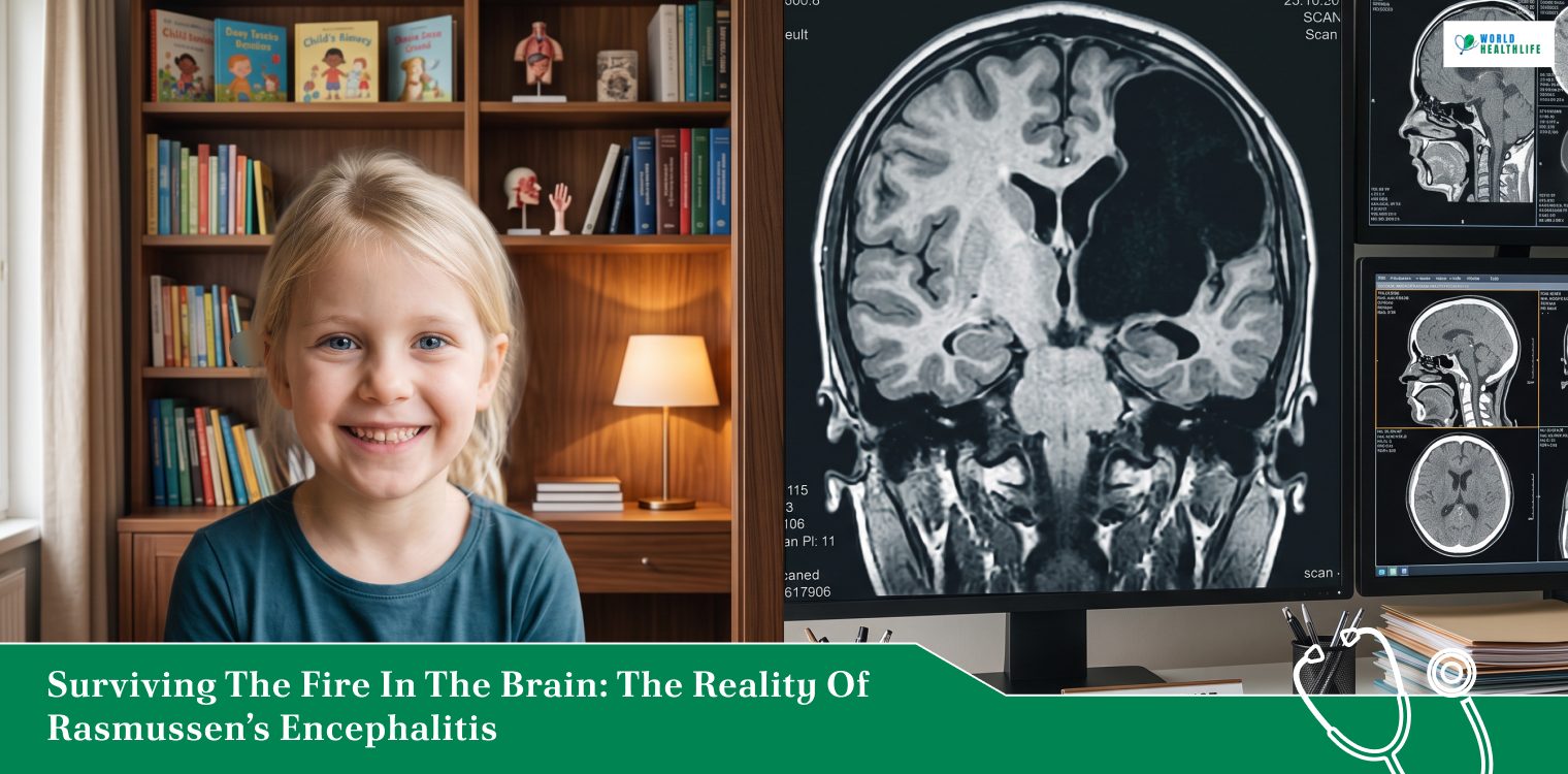
Imagine a medical crisis such as this one.
A child’s brain goes to war with itself, systematically dismantling an entire hemisphere while standard medicines stand by completely helpless.
This is not a hypothetical worst-case scenario. Conversely, it is the daily reality for families navigating Rasmussen encephalitis, an extraordinarily rare, aggressive inflammatory neurological condition.
First identified by neurosurgeon Theodore Rasmussen in 1958, this disease primarily targets children under the age of ten.
Acting like a slow-moving localized fire, the condition causes progressive brain shrinking, cognitive decline, and relentless seizures. Furthermore, these seizures are resistant to any form of treatment.
Because it strikes without warning and causes rapid, irreversible neurological damage, understanding its progression and treatment landscape is absolutely vital for clinical teams and caregivers alike.
Thus, the following guide will delve deep into the whole reality of the disease, how it evolves, clinical signs, diagnosis, and treatment plans.
The Path Of Destruction: How The Disease Evolves
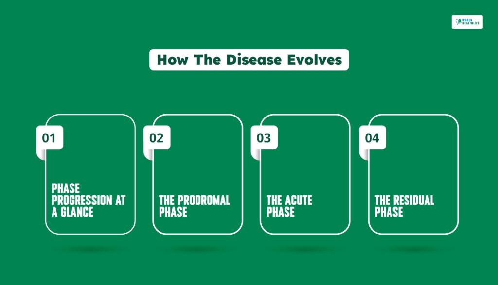
Rasmussen encephalitis does not destroy the brain all at once. Instead, it moves through three brutal, distinct clinical phases. Gradually, the condition claims territory in one half of the brain.
Phase Progression At A Glance
| Disease Phase | Typical Duration | Primary Defining Characteristic |
| Prodromal | Months to several years | Mild, infrequent focal seizures and subtle clumsiness. |
| Acute | 8 to 12 months | Peak inflammation, constant seizures (EPC), and rapid paralysis. |
| Residual | Permanent / Lifelong | Burned-out inflammation with fixed, severe neurological deficits. |
The Prodromal Phase
In the beginning, the warning signs are subtle and easily missed. A child might experience occasional isolate focal seizures or slight clumsiness on one side of the body.
This stage can last anywhere from a few months to several years, masking the true severity of what is to come.
The Acute Phase
Usually lasting between 8 and 12 months, this is considered one of the most destructive windows.
The underlying inflammation reaches its peak, triggering near-constant intractable seizures.
Many patients develop a terrifying condition known as Epilepsia Partialis Continua (EPC). Here, a specific muscle group, such as a hand or half the face, jerks in a rhythmic manner, which can be continuously for hours or even days on end.
Physical weakness rapidly worsens into partial paralysis, where cognitive development often grinds to a halt.
The Residual Phase
Eventually, the active inflammatory fire burns itself out. However, the structural damage does permanent harm.
The affected hemisphere is left severely scarred and shrunken. While the continuous seizures may finally slow down, the illness leaves the patient with fixed and profound neurological defects.
These also include permanent weakness as well as cognitive challenges.
Mapping The Clinical Stages
To separate this condition from other forms of epilepsy, neurologists look for a specific cluster of asymmetric symptoms.
The table below details how the disease alters function over time.
| Affected Area | Clinical Presentation | Long-Term Impact |
| Motor Control | Progressive hemiparesis (weakness on one side of the body). | Eventually leads to complete hemiplegia (paralysis on one side). |
| Seizure Profile | Simple focal seizures that rapidly escalate to continuous EPC. | Complete resistance to standard anti-epileptic medications. |
| Vision & Language | Loss of the same half of the visual field in both eyes. | Severe loss of speech (aphasia) if the dominant hemisphere is attacked. |
| Cognition | Accelerated memory loss and executive function decline. | Long-term learning disabilities and developmental arrest. |
Finding the Visual Evidence: Diagnosis and MRI Changes
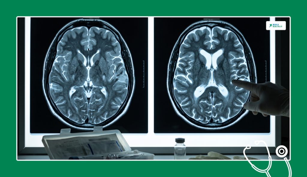
Catching this disease early requires careful tracking through serial magnetic resonance imaging (MRI) scans. In the earliest stages, a brain scan might look completely normal or show only faint swelling in isolated spots.
However, as the months roll by, serial MRIs reveal a striking, unmistakable diagnostic hallmark: progressive unilateral hemispheric atrophy.
One side of the brain visibly shrinks away. As a result, this leaves the fluid-filled cavities on that side vastly enlarged. On the other hand, the opposite side of the brain remains completely untouched.
Doctors also utilize lumbar punctures to check for elevated inflammatory markers in the cerebrospinal fluid, confirming that an immune response is driving the damage.
Treatment Strategies: From Medication to Disconnection
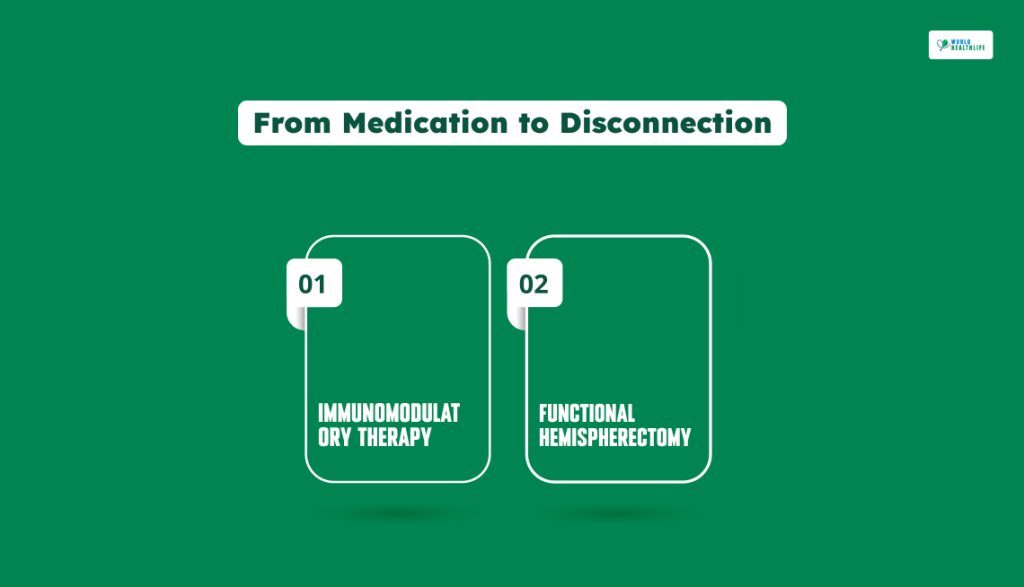
There is no cure for Rasmussen encephalitis. The reason behind this is that standard anti-seizure drugs almost completely fail to control the episodes.
Additionally, medical teams must pivot to aggressive interventions to slow the damage and preserve the quality of life.
Immunomodulatory Therapy
In order to dampen the immune system’s attack on the brain, doctors deploy high-dose steroids, intravenous immunoglobulin (IVIG), and plasmapheresis.
As a result, this filters the blood.
Doctors also use immunosuppressive medications to prolong the acute phase and delay tissue loss, though they rarely stop the disease permanently.
Functional Hemispherectomy
When medical options fail, surgery becomes the definitive line of defense.
A functional hemispherectomy involves surgically disconnecting the diseased, non-functional half of the brain from the healthy half. Furthermore, it leaves the physical tissue in place.
While this operation permanently sacrifices fine motor control on one side of the body and alters peripheral vision, it delivers an astonishingly high rate of complete seizure freedom.
For young children, the remaining healthy half of the brain displays incredible neuroplasticity. In addition, it often rewires itself to take over language and cognitive tasks entirely.
Navigating The Diagnosis: Specialized Support And Resources
Coping with a rare, progressive condition like Rasmussen encephalitis places an immense emotional, financial, and educational strain on the entire family unit.
Medical management often moves toward complex surgical decisions. As a result, connecting with specialized advocacy networks can provide families with vital guidance and community peer support.
| Organization Name | Primary Strategic Focus | Key Resource or Peer Inclusions |
| The Hemispherectomy Foundation | Practical and financial aid for families facing or recovering from radical brain surgery. | Provides financial assistance alongside active community groups, including the Rasmussen’s Encephalitis Family Network peer forum. |
| Pediatric Epilepsy Surgery Alliance (PESA) | Educational advocacy and parent-matching support pipelines. | Offers downloadable family guides, specialized webinars, and structured training materials tailored for a child’s school education team. |
| RE Children’s Project | Funding scientific research and driving broad clinical disease awareness. | Aggressively finances medical studies targeting post-surgical brain recovery timelines and ultimate cure discovery initiatives. |
| Encephalitis International | Global clinical outreach and mental health support. | Hosts regular virtual peer support groups via video platforms and provides direct access to localized counseling directories. |
Facing The Future
Dealing with this diagnosis requires shifting expectations from a standard medical cure to long-term adaptation.
The journey is incredibly difficult, forcing families to accept a trade-off between controlled seizures and permanent physical deficits.
Yet, the human brain’s ability to adapt and rebuild after a hemispherectomy is a testament to neurological resilience.
By combining early detection with aggressive surgical timing, clinical teams can successfully extinguish the electrical storms, giving children a chance to rebuild their lives with a single, healthy hemisphere.
Disclaimer: The information provided in this article is for general informational purposes only. It does not, and is not intended to, constitute medical or health advice. Please consult a qualified healthcare professional for medical guidance or expert health assistance.
References
Khan Z, Arya K, Bollu PC. Epilepsia Partialis Continua. [Updated 2023 Aug 28]. In: StatPearls [Internet]. Treasure Island (FL): StatPearls Publishing; 2026 Jan.


