The structural framework of the human brain develops through a highly complex and microscopic choreography during early pregnancy.
Specifically, between the second and fifth months of gestation, billions of primitive neurons travel outward from the deep ventricular zones. These then assemble the outer cerebral cortex.
This process must work perfectly for the creation of a unified and functional computing engine.
However, sometimes, a severe structural breakdown halts this structural timeline mid-construction. This happens in one such rare congenital disorder of neuronal migration.
In cases of schizencephaly, the wall of the brain hemisphere fails to close completely. Instead, an abnormal and fluid-filled structural cleft cuts all the way through the brain tissue.
Moreover, it stretches from the deep inner fluid cavities, or the lateral ventricles, outward to the surface grey matter.
This ultra-rare structural brain malformation presents a massive lifelong neurological hurdle.
Because the cleft interrupts normal motor pathways and shorts out grey matter connectivity, children born with this condition face severe focal seizures, developmental arrest, and hemiparesis.
Therefore, managing life with this diagnosis requires a raw, realistic look at its physical structural tiers, its developmental impact, and the core therapeutic pipelines.
Defining The Structural Defect: Closed-Lip Vs. Open-Lip Architecture
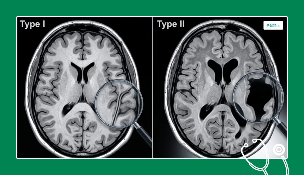
Medical professionals divide the disorder into two starkly different structural types based on the physical configuration of the brain cleft.
As a rule, the alignment of the grey matter walls completely dictates how severe the physical tracking symptoms will be.
Type I: Closed-Lip Schizencephaly
In the closed-lip variant, the structural cleft inside the hemisphere remains narrow and compressed. The grey matter walls lining the cleft physically touch one another, leaving no open space.
On a diagnostic screen, this looks like a thin, dark line tracking through the white matter. Because the surrounding brain tissue remains structurally supported, Type I cases typically present with milder symptoms.
For instance, these include localized muscle weakness or easily managed focal seizures.
Type II: Open-Lip Schizencephaly
On the other hand, the open-lip variant is significantly more destructive. Here, the grey matter walls are separated wide apart, leaving a physical void in the hemisphere.
Cerebrospinal fluid (CSF) fills this gap directly from the ventricles, creating a large, fluid-filled channel that bypasses normal brain structures.
Type II cases frequently trigger high intracranial pressure, progressive macrocephaly, and a severe condition called hydrocephalus.
Because a large chunk of functional brain tissue is missing, the clinical complications are far more aggressive.
The Structural Severity Spectrum
The physical reach of the structural cleft determines a child’s exact physical limitations.
Clinicians evaluate whether the defect impacts one or both sides of the brain to map out the long-term prognosis.
| Severity Tier | Anatomical Presentation | Core Clinical Footprint | Long-Term Mobility Outlook |
| Unilateral Closed-Lip | A single, tight cleft localized to one hemisphere. | Mild hemiparesis, normal head circumference, and highly responsive epilepsy. | Children frequently learn to walk independently and live with near-normal cognitive function. |
| Unilateral Open-Lip | A wide, fluid-filled structural void in one side of the brain. | Moderate motor delays, visible one-sided body weakness, and frequent focal seizures. | Requires focused physical therapy and braces; cognitive outcomes vary from mild to moderate delay. |
| Bilateral Closed-Lip | Narrow, touching clefts cutting through both hemispheres. | Significant spastic diplegia, delayed motor milestones, and global learning hurdles. | Walking typically requires walking frames or rolling chairs; requires lifelong support. |
| Bilateral Open-Lip | Wide, open structural voids destroying large zones on both sides of the brain. | Catastrophic epilepsy, developmental arrest, severe microcephaly, or hydrocephalus. | Children remain completely non-ambulatory, requiring intensive around-the-clock physical care. |
Unraveling The Causes: Genetic Triggers And Vascular Insults
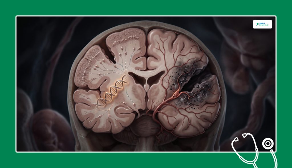
Pinpointing the exact cause of a brain cleft remains a difficult task for geneticists. The condition is highly heterogenous.
This means it can stem from different biological pathways during early fetal development.
The EMX2 Gene Mutation
A small percentage of familial cases are driven by specific mutations in the EMX2 homeobox gene.
Additionally, this gene acts as a structural foreman during pregnancy, providing the instructions to organize the boundaries of the developing cerebral cortex.
When a mutation disrupts this signaling pathway, the cell migration line breaks down entirely. The neurons fail to bridge the gaps, leaving an unclosed structural cleft behind.
Intrauterine Vascular Insults
The majority of cases happen randomly and are not inherited from parents.
The leading medical theory points toward a localized vascular disruption, occurring within the fetal brain around the seventh or eight week of gestation. This disruption is a mini-stroke or a sudden loss of blood flow.
If the middle cerebral artery system experiences a temporary blockage, the young cells in that specific zone suffocate and die.
The surrounding healthy cells continue to grow and migrate normally, leaving a physical structural gap where the tissue died.
As a result, this localized destruction can also be triggered by congenital intrauterine infections, such as cytomegalovirus (CMV).
Associated Midline Brain Anomalies
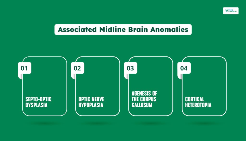
A brain cleft rarely happens in total isolation. Since the early cellular error impacts the entire midline blueprint of the central nervous system, radiologists frequently spot a cluster of co-occurring structural malformations.
- Septo-Optic Dysplasia: The complete or partial absence of the septum pellucidum, the thin membrane separating the brain’s fluid cavities. Coupled with underdeveloped optic nerves.
- Optic Nerve Hypoplasia: Abnormally small, poorly wired optic nerves that lead to severe, permanent visual impairments.
- Agenesis of the Corpus Callosum: The missing connection bridge between the left and right hemispheres, which severely limits cross-brain communication.
- Cortical Heterotopia: Trapped clusters of grey matter cells that got stuck deep inside the white matter tracts during their outward migration journey.
Diagnostic Confirmations: The Superiority Of High-Resolution MRI
Catching the condition early requires advanced neuroimaging.
While a prenatal ultrasound can occasionally spot a wide and open-lip void in late pregnancy, a postnatal magnetic resonance imaging (MRI) scan is the gold standard for confirmation.
Neuroradiologists rely on one absolute rule to separate schizencephaly from other destructive conditions like porencephaly.
That is, the cleft must be completely lined with grey matter. Porencephaly is just a dead fluid cavity lined with scarred white matter tissue.
The presence of heterotopic grey matter running along the entire edge of the channel proves one thing. Instead of just a random late-pregnancy injury, the issue was a fundamental failure of cellular migration.
Additionally, the MRI tracks ventricular dilation, helping neurosurgeons monitor for early signs of hydrocephalus.
Spotting The Complications: Clinical Presentation And Symptoms
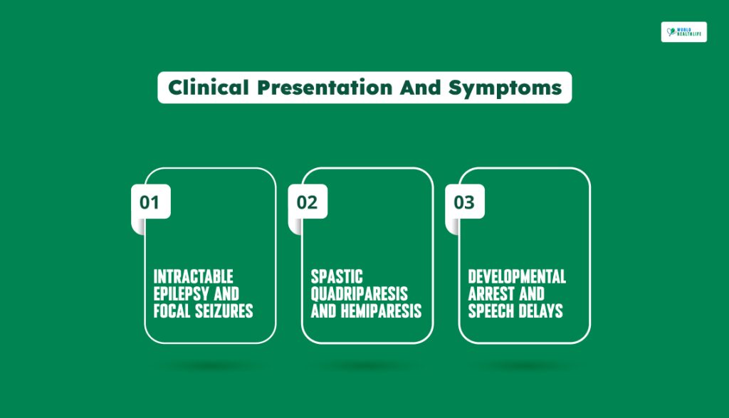
Since a structural cleft disrupts the brain’s internal wiring, it triggers a predictable wave of physical and cognitive hurdles very early in life.
Intractable Epilepsy And Focal Seizures
Erratic electrical storms are the biggest daily hurdle.
Over 80% of patients develop epilepsy, which usually manifests as focal seizures targeting one specific limb or one side of the face.
In severe bilateral cases, these episodes can escalate into infantile spasms or generalized tonic-clonic seizures that are highly resistant to standard medications.
Spastic Quadriparesis And Hemiparesis
The motor tracts coming down from the cortex are physically cut in half by the cleft.
Children with unilateral defects experience hemiparesis, leaving one side of the body stiff and weak.
Those with bilateral open-lip defects deal with severe spastic quadriparesis, profound stiffness, and near-total paralysis across all four limbs.
Developmental Arrest and Speech Delays
When clefts destroy tissue on both sides of the brain, early milestones like tracking objects, babbling, and rolling over grind to a halt.
In addition, language processing is heavily impacted if the cleft cuts through the dominant hemisphere, leading to severe dysarthria or full mutism.
Navigating The Care And Treatment Pipeline
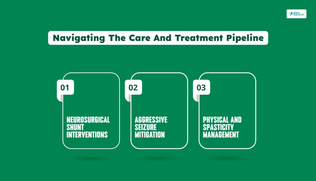
There is no way to surgically patch an open brain cleft or restart fetal cell migration after birth.
A direct cure does not exist. As a result, medical teams focus entirely on managing secondary brain pressures, soothing rigid muscle configurations, and controlling seizures.
Neurosurgical Shunt Interventions
If an open-lip cleft triggers hydrocephalus, a neurosurgeon must act quickly. They will surgically place a ventriculoperitoneal (VP) shunt to drain excess fluid out of the brain cavities.
Consequently, this relieves dangerous intracranial pressure and prevents further tissue loss.
Aggressive Seizure Mitigation
Neurologists utilize combinations of anti-epileptic medications to stabilize the brain’s chaotic chemistry.
If standard drugs fail, families often implement a strict, medically managed ketogenic diet to naturally lower seizure frequencies.
Physical And Spasticity Management
Continuous physical and occupational therapy is essential to prevent rigid limbs from locking into permanent, painful shapes called contractures.
Physicians use custom back braces, standing frames, and targeted botulinum toxin (Botox) injections to temporarily relax stiff muscle groups and preserve joint mobility.
Strategic Long-Term Assessment
A diagnosis fundamentally shifts a family’s journey, replacing standard milestone charts with an intense focus on comfort, mobility, and symptom management.
Lifespan outcomes vary wildly. Children with small, unilateral closed clefts can live completely full, near-independent lives.
Those facing bilateral open-lip configurations require intensive, around-the-clock clinical care to manage chronic infections and respiratory vulnerabilities.
You cannot find progress on a standard growth scale.
Instead, you find it in the quiet, significant victories. It can be a day completely free from the exhaustion of seizures, a comfortable feeding session, or a successful transition into an adaptive communication system.
A focus on aggressive comfort care and early therapeutic support can allow clinical teams to help these families navigate this complex structural architecture. Additionally, they can do so with immense dignity and long-term resilience.
Disclaimer: The information provided in this article is for general informational purposes only. It does not, and is not intended to, constitute medical or health advice. Please consult a qualified healthcare professional for medical guidance or expert health assistance.