
When you discover that you have a growth inside an internal organ, it can be quite an unsettling experience.
However, learning about gallbladder polyps is actually the first step that you take towards understanding your digestive health.
First of all, let us understand one thing. Your gallbladder.
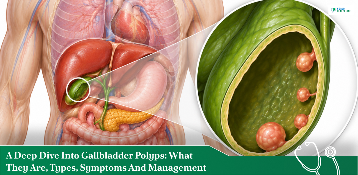
This is a small, pear-shaped organ that is nestled beneath your liver and stores and concentrates bile. Moreover, bile is a vital fluid required for breaking down dietary fats.
When small, elevated mucosal lesions or cellular growths project outward from the inner lining of this organ’s wall, they are medically classified as polyps.
Approximately 95% of these growths are completely benign, which is noncancerous. Moreover, they rarely spark noticeable discomfort.
So, most individuals only discover they have them incidentally during routine medical imaging for entirely unrelated abdominal complaints.
However, navigating the diagnosis requires distinguishing between harmless tissue clusters and true cellular growths. These clusters carry a small risk of developing into malignancy over time.
So, in this guide, we are taking a closer look at the underlying causes, common risk factors, structural variations, and tracking protocols.
In this way, you can work closely with your healthcare provider to manage your condition safely and effectively.
What Are Gallbladder Polyps

Let us understand gallbladder polyps in a deeper way.
So, gallbladder polyps are elevated growths or lesions that project outward from the inner mucosal lining into the lumen, where bile is stored. Here, the lumen is the interior cavity.
Affecting roughly 4% to 7% of the general population, these are highly common.
In addition, they are most frequently discovered incidentally during routine abdominal ultrasounds performed for unrelated digestive complaints.
Approximately 95% of gallbladder polyps are completely benign. Clinically, these are categorized into two primary classifications.
- Pseudopolyps or non-neoplastic lesions
- True polyps or neoplastic lesions
Types Of Gallbladder Polyps
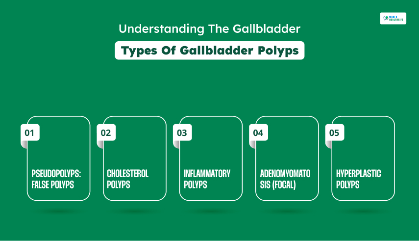
While the majority of these growths are completely harmless, they are clinically categorized into three major types based on two things. Their cellular makeup and their risk of progression.
At A Glance: Comparing The Main Types
| Feature | Pseudopolyps (False) | True Polyps (Benign) | Malignant Polyps (Cancerous) |
| Frequency | 60% – 90% (Very Common) | Uncommon | ~5% (Rare) |
| Cancer Risk | None | Precancerous (Can transform) | Is Cancerous |
| Typical Size | Usually < 10 mm | Variable, risk increases if > 10 mm | Frequently > 10 mm (often > 18 mm) |
| Quantity | Frequently multiple | Usually solitary | Usually solitary |
Pseudopolyps: False Polyps
Pseudopolyps are actually not true tumors. Instead, they represent approximately 60% to 90% of all diagnosed gallbladder polyps while possessing zero malignant potential.
Instead of arising from autonomous cellular replication, they arise from:
- Metabolic deposits
- Chronic inflammation
- Structural changes
| Subtype | Primary Features & Pathophysiology |
| Cholesterol Polyps | • 60-90% of cases • Caused by lipid buildup • Multiple, small (< 5 mm) • Yellowish • Fragile stalks |
| Inflammatory Polyps | • 5–10% of cases • Driven by chronic cholecystitis • Fibrous tissue and immune cell infiltration |
| Adenomyomatosis (Focal) | • Epithelial invagination into the muscular wall • Creates Rokitansky-Aschoff sinuses; “comet-tail” |
| Hyperplastic Polyps | • Rare • Localized overgrowth of normal mucosa • Completely benign structural variations |
Cholesterol Polyps
In the case of cholesterol polyps, their formation happens due to a localized defect in cholesterol metabolism.
Excess cholesterol crystals deposit within the lamina propria of the gallbladder wall. Moreover, immune cells known as macrophages engulf these lipids, transforming into lipid-laden “foam cells.”
Typically, they present as multiple, small, yellowish, fragile lesions that are attached to the mucosa by a thin, delicate pedicle (stalk).
In addition, they can easily detach, occasionally contributing to biliary colic if they block the cystic duct.
Inflammatory Polyps
They are a direct consequence of mechanical irritation or persistent low-grade infections.
Generally, these types of polyps develop from chronic inflammation (chronic cholecystitis) and are highly correlated with the presence of gallstones (cholelithiasis).
When it comes to its composition, it is a mixture of fibrous scar tissue, granulation tissue, and a dense infiltration of inflammatory immune cells.
These cells are plasma cells and lymphocytes.
They are typically sessile (flat-based) or irregular in shape.
Adenomyomatosis (Focal)
This type of polyp is characterized by excessive proliferation of the surface epithelium alongside hypertrophy (thickening) of the smooth muscular layer of the gallbladder wall.
This overgrowth forces the mucosal lining to invaginate outward into the muscular layer. As a result, this creates deep diverticula known as Rokitansky-Aschoff sinuses.
Technically, this is a structural wall abnormality which is also termed hyperplastic cholecystosis.
However, instead of a discrete polyp, a focal presentation at the fundus of the gallbladder frequently mimics a polyp on a transabdominal ultrasound.
This often produces a classic “comet-tail” artifact.
Hyperplastic Polyps
These are rare and completely benign lesions that occur when normal mucosal epithelial cells multiply slightly faster than usual.
As a result, this leads to a small, localized fold or bump. Moreover, they do not demonstrate cellular dysplasia, which is abnormal development, or atypia.
True Polyps: Neoplastic and Benign Tumors
True benign polyps are genuine cellular neoplasms that result from autonomous, abnormal multiplication of the epithelial or mesenchymal cells lining the gallbladder.
While benign at the time of discovery, certain subtypes require strict surveillance due to their potential to evolve over time.
Adenomas
Adenomas can be defined as true epithelial tumors that are composed of glandular tissue.
Representing a critical clinical entity due to being inherently precancerous, they progress through a well-documented adenoma-carcinoma sequence.
Here, genetic mutations accumulate over a period of years. Eventually, they transform benign glandular tissue into invasive malignancy.
Adenomas can be tubular, papillary, or tubulopapillary. Additionally, they are typically larger than pseudopolyps, exceeding 10mm.
Besides that, they are solitary and can be either pedunculated on a stalk or sessile.
Malignant Polyps: Cancerous
Malignant polyps represent aggressive, primary gallbladder cancers presenting as intraluminal masses.
In addition, they account for approximately 5% of all detected gallbladder polyps.
Consequently, accurate differentiation is a medical priority because gallbladder malignancies are highly aggressive.
Adenocarcinomas
The predominant histological subtype of gallbladder cancer is adenocarcinoma.
They arise directly from the glandular epithelial cells, where early-stage adenocarcinomas often present as small, localized mucosal projections.
On a standard transabdominal ultrasound, a small, early-stage adenocarcinoma can look completely identical to a harmless cholesterol polyp or a benign adenoma.
This makes tracking specific structural characteristics essential.
Malignant Features
| Features | Description |
| Rapid Growth Rate | • Fast • Progressive increases in size noted during serial follow-up imaging • Strongly indicate active malignancy |
| Solitary Presentation | • Different from cholesterol polyps • Are always almost single isolated lesions |
| Sessile Morphology | • Broad and flat base (sessile) • Higher likelihood of malignancy compared to a narrow and stalked presentation |
| Wall/Tissue Invasion | • Ultrasound, CT, or MRI evidence shows loss of the normal gallbladder wall layers • Direct tissue invasion into the surrounding liver bed • Definitive indicator of an advanced malignant tumor |
Causes Of Gallbladder Polyps
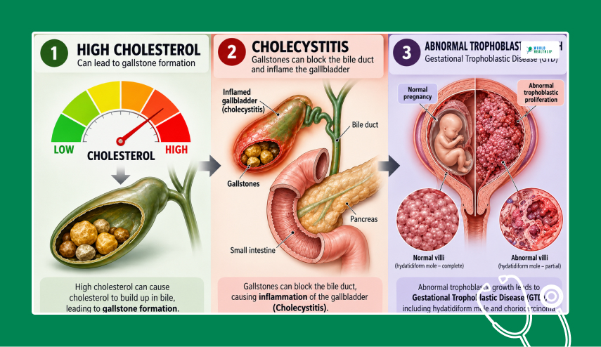
The primary causes of gallbladder polyps include the buildup of cholesterol, chronic inflammation, or abnormal cell growth in the gallbladder wall.
Cholesterol Buildup: Cholesterolosis
In cholesterolosis, there is an excess buildup of cholesterol or bile salts that can form crystals that immune cells or macrophages engulf.
Here, these fat-filled cells stick to the inner wall of the gallbladder. As a result, this leads to the creation of harmless cholesterol pseudopolyps.
This is also termed “strawberry gallbladder”
Chronic Inflammation: Cholecystitis
In this case, long-term irritation or repeated injury from gallstones or infections causes scar tissue and inflammatory cells to accumulate.
This forms inflammatory polyps.
Abnormal Tissue Proliferation
In the case of true polyps such as adenomas or adenomyomatosis, they develop in another way.
That is, when the surface epithelium or glandular cells of the gallbladder wall grow, thicken, or form small pouches or invaginations.
Associated Risk Factors For Gallbladder Polyps

| Risk Factor Category | Specific Factors & Conditions | Impact / Clinical Presentation |
| Local Irritation | Gallstones | Frequently co-exist with polypoid lesions or contribute directly to localized tissue irritation in the gallbladder lining. |
| Infections | Chronic viral infections (e.g., Hepatitis B) | Long-term viral infections have been clinically linked to an increased risk of abnormal tissue and polyp formation. |
| Genetic & Inherited Syndromes | • Familial Adenomatous Polyposis (FAP) • Peutz-Jeghers syndrome • Gardner syndrome | Systemic conditions that cause gastrointestinal polyps substantially raise the underlying likelihood of developing gallbladder growths. |
| Bile Duct Conditions | Primary Sclerosing Cholangitis (PSC) | Chronic inflammatory diseases of the bile ducts significantly increase the vulnerability to developing gallbladder lesions. |
| Demographics & Metabolism | • Age (particularly over 50–60 years) • Obesity • High blood lipid levels | Metabolic factors and advanced age are the most frequently observed clinical characteristics in individuals diagnosed with gallbladder polyps. |
Common Symptoms Of Gallbladder Polyps
Gallbladder polyps themselves rarely cause any symptoms.
Instead, in approximately 90% to 95% of cases, they are completely asymptomatic.
Usually, they are discovered entirely by accident when an individual undergoes an abdominal ultrasound or CT scan for an unrelated medical reason.
When symptoms do start to occur, they are typically not caused directly by the polyp tissue itself.
Rather, they occur by the polyp interfering with normal bile flow or coexisting with other gallbladder conditions such as gallstones or chronic inflammation.
| Presentation | Associated Clinical Finding | Typical Next Steps |
| Completely Asymptomatic | Small pseudopolyps or early-stage benign growths (<5–6 mm) | Standard monitoring via serial ultrasounds at fixed intervals; no immediate surgery needed. |
| Intermittent Right Upper Quadrant Pain | Polyp fragment blocking bile flow or concurrent gallstones | Evaluation for a cholecystectomy (gallbladder removal) to relieve chronic irritation. |
| Jaundice, Fever, or Severe Pain | Acute bile duct obstruction or advanced tissue changes | Urgent diagnostic imaging and surgical consultation to prevent systemic complications. |
How Do Doctors Diagnose Gallbladder Polyps

Gallbladder polyps do not usually cause symptoms on their own. Due to this, doctors almost always diagnose them incidentally while looking for other conditions.
For example, gallstones or general abdominal pain are other conditions.
When a physician suspects or detects a polyp, they rely on specific imaging modalities to do the following:
- Confirm its presence
- Measure its size
- Evaluate its potential for malignancy
Diagnostic Workup Overview
| Imaging Modality | Clinical Role | What it Identifies |
| Transabdominal Ultrasound | First-line choice; standard screening tool | Confirms presence; differentiates fixed polyps from mobile gallstones |
| Endoscopic Ultrasound (EUS) | Advanced evaluation of borderline polyps (6–9 mm) | High-resolution wall layering; checks for micro-invasion or malignant features |
| CT / MRI (MRCP) | Staging and complex case evaluation (>10 mm) | Assesses liver involvement, lymph node spread, or associated bile duct conditions |
Transabdominal Ultrasound: First-Line Diagnostic Tool
Doctors consider the abdominal ultrasound the gold standard for diagnosing gallbladder polyps.
Moreover, it is highly sensitive, non-invasive, and cost-effective.
Let us understand how this works.
Here, high-frequency sound waves create real-time images of the gallbladder. A polyp appears as a fixed, bright projection that extends from the wall into the fluid-filled interior.
Unlike gallstones, polyps do not move when the patient changes positions and do not cast an acoustic shadow behind them.
Endoscopic Ultrasound (EUS)
There are times when a transabdominal ultrasound fails to reveal a borderline polyp, which is typically 6 mm to 9mm.
Moreover, it may also be that the doctor cannot tell if a mass is benign or malignant. In this case, the doctor performs an endoscopic ultrasound (EUS).
This procedure involves passing a thin, flexible tube with a tiny ultrasound probe at its tip down into the stomach.
Following this, you position it directly next to the gallbladder.
Because the probe is physically closer to the organ, it provides incredibly high-resolution, detailed images of the individual layers of the gallbladder wall.
Thus, clinicians can differentiate between harmless cholesterol polyps, precancerous adenomas, and early-stage cancer.
Computed Tomography (CT) Scan
Doctors generally do not use a standard Computed Tomography (CT) scan or Magnetic Resonance Imaging (MRI) for initial diagnosis.
This is because they can easily miss lesions smaller than 10 mm.
However, they become critical if a polyp is larger than 10 mm or displays suspicious features.
Doctors utilize contrast-enhanced CT scans to check if a suspected tumor has grown into the liver wall or spread to nearby lymph nodes.
Magnetic Resonance Cholangiopancreatography (MRCP)
On the contrary, doctors use a specialized MRI called an MRCP (Magnetic Resonance Cholangiopancreatography) to visualize the surrounding bile ducts.
Doctors also use them to check for concurrent issues like Primary Sclerosing Cholangitis.
Treatment And Management
Polyp size and specific structural characteristics heavily dictate the clinical management of gallbladder polyps through risk stratification.
Since it is impossible to definitively differentiate between a benign adenoma and an early-stage malignancy using standard imaging alone, physicians use objective measurement thresholds.
This helps them to decide between conservative monitoring and surgical intervention.
Risk Stratification
| Polyp Size / Characteristic | Risk Assessment | Recommended Clinical Actions |
| Smaller than 5 mm | Very low to negligible risk of malignancy. | Routine surveillance is rarely required unless patient risk factors are elevated. |
| 6 mm to 9 mm | Low risk; primarily monitored for active growth. | Ultrasound surveillance at regular intervals (typically 6 months, 1 year, and 2 years) to track changes. |
| 10 mm (1 cm) or larger | High risk of malignancy or malignant transformation. | Cholecystectomy (surgical removal of the gallbladder), usually performed laparoscopically. |
| Rapid Growth | Highly suspicious if a polyp grows by 2 mm or more between consecutive scans. | Prompt surgical removal of the gallbladder. |
Disclaimer: The information provided in this article is for general informational purposes only. It does not, and is not intended to, constitute medical or health advice. Please consult a qualified healthcare professional for medical guidance or expert health assistance.
References
- Weerakkody, Y., Di Muzio, B., & Knipe, H. (2009). Gallbladder polyp. Radiopaedia.org.
- Kim, K. H. (2020). Gallbladder polyps: evolving approach to the diagnosis and management. Journal of Yeungnam Medical Science, 38(1), 1–9. Yeungnam University Institute of Medical Science.
- Jones, M. W., & Deppen, J. G. (2023). Gallbladder polyp. StatPearls – NCBI Bookshelf.
- Kamaya, A., Fung, C., Szpakowski, J., Fetzer, D. T., Walsh, A. J., Alimi, Y., Bingham, D. B., Corwin, M. T., Dahiya, N., Gabriel, H., Park, W. G., Porembka, M. R., Rodgers, S. K., Tublin, M. E., Yuan, X., Zhang, Y., & Middleton, W. D. (2022). Management of incidentally Detected Gallbladder Polyps: Society of Radiologists in Ultrasound Consensus Conference Recommendations. Radiology, 305(2), 277–289. Radiological Society of North America.
- Dilek, O. N., Karasu, S., & Dilek, F. H. (2019). Diagnosis and Treatment of Gallbladder Polyps: Current Perspectives. European Journal of Hepato-Gastroenterology, 9(1), 40. National Library of Medicine.
- Cleveland Clinic. (2026). Gallbladder polyps. Cleveland Clinic.




What to Eat After Gallbladder Surgery: Ultimate Recovery Diet
October 30, 2026[…] small polyps are generally considered harmless, any mucosal growth that reaches 10 mm or larger, or a rapid […]